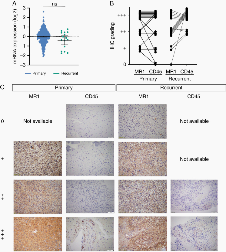
Figure 3.
MR1 and CD45 expression does not vary between primary and recurrent glioblastoma. (A) TCGA data show no significant difference between primary and recurrent tumors MR1 mRNA expression levels in GBM. (B) Quantification of MR1 and CD45 IHC staining levels in matched primary and recurrent GBM tissues. (C) IHC of primary and recurrent tumors that were stained for MR1 and CD45 expression. Staining levels for MR1: 0, not detected; +, low (33%); ++, medium (66%); +++, high (˃66%). For CD45 tissue was divided into 4 quadrants: 0 non-detected, +: 1 quadrant positive, ++: 2 quadrants positive, and +++: ˃2 quadrants positive.

