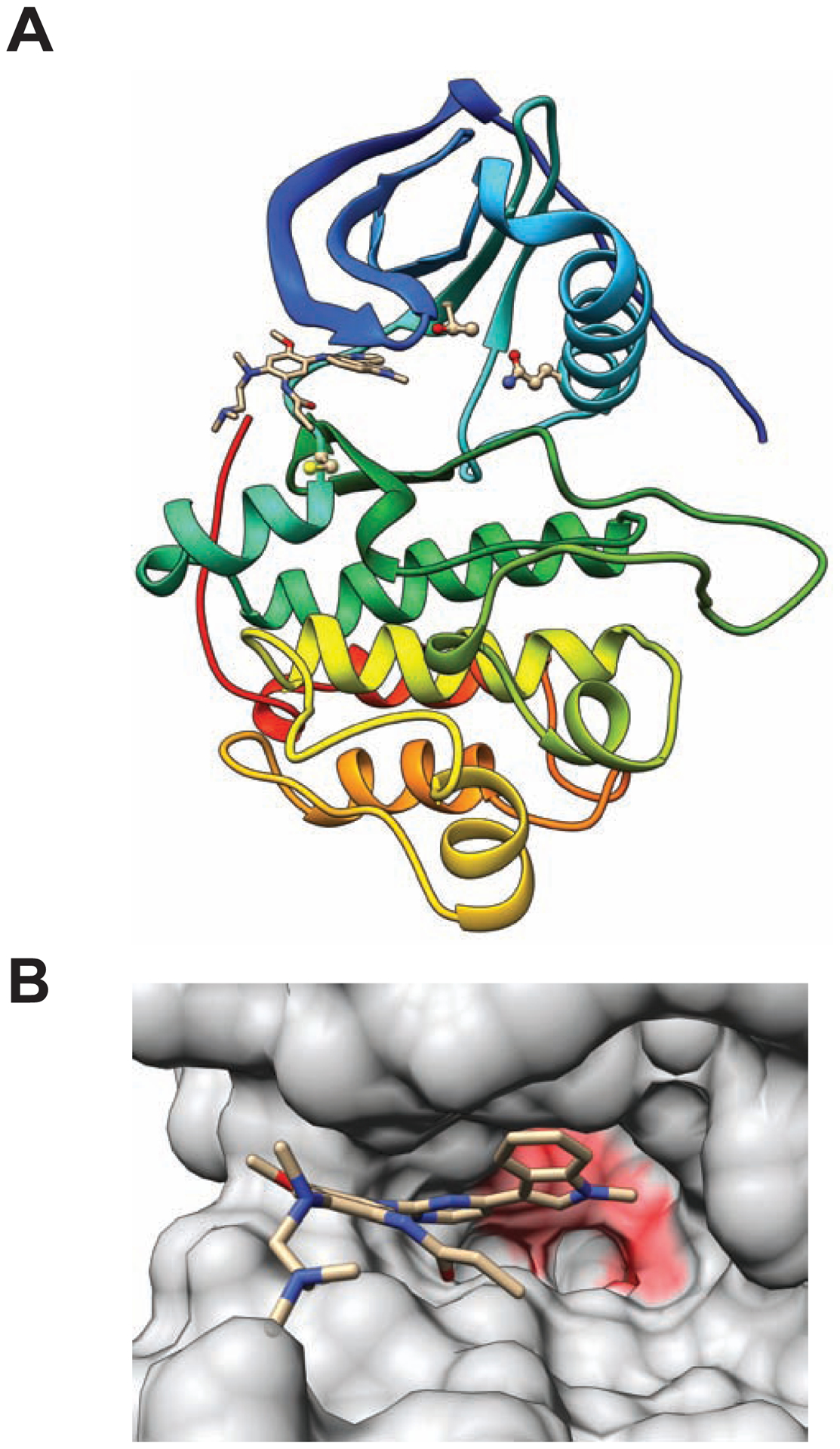
Figure 1.

Homology models of EGFR L858R/M766Q double mutant with bound osimertinib. (A) Protein-ligand complex structure. Protein chain shown with cartoon representation; inhibitor shown with stick figure representation; and residues Q766, T790, and C797 shown with ball and stick representations. (B) Inhibitor binding site. Solvent-accessible protein surface is shown with semi-transparent representation. Osimertinib is shown with stick representation. Surface features overlying residues with predicted unfavorable steric clashes are colored red.
