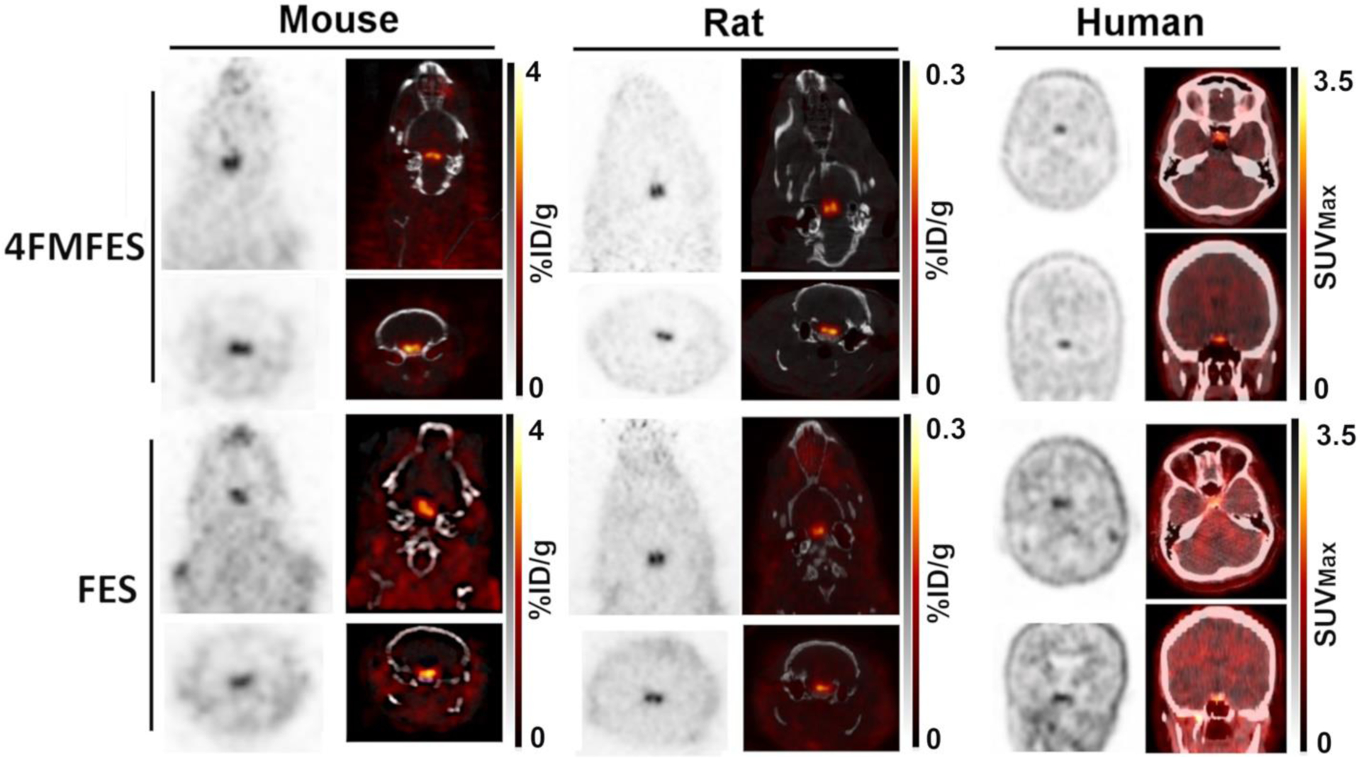
Figure 1: Cross-species 4FMFES- and FES-PET brain imaging.

Representative coronal and transaxial images of brain slices from 4FMFES and FES PET/CT for mouse (left panels), rat (middle panels) and human (right panels), all centered on the pituitary. The patient shown was a 57 years-old post-menopausal woman. The grayscale (PET) and hot metal scale (PET component in the PET/CT fusion images) uptake values are indicated for each dataset in %ID/g for animal studies and in SUVMax for clinical images. Rodent images were obtained on the LabPET8/Triumph preclinical PET/CT platform. Clinical images were obtained on a Philips TF whole-body PET/CT scanner.
