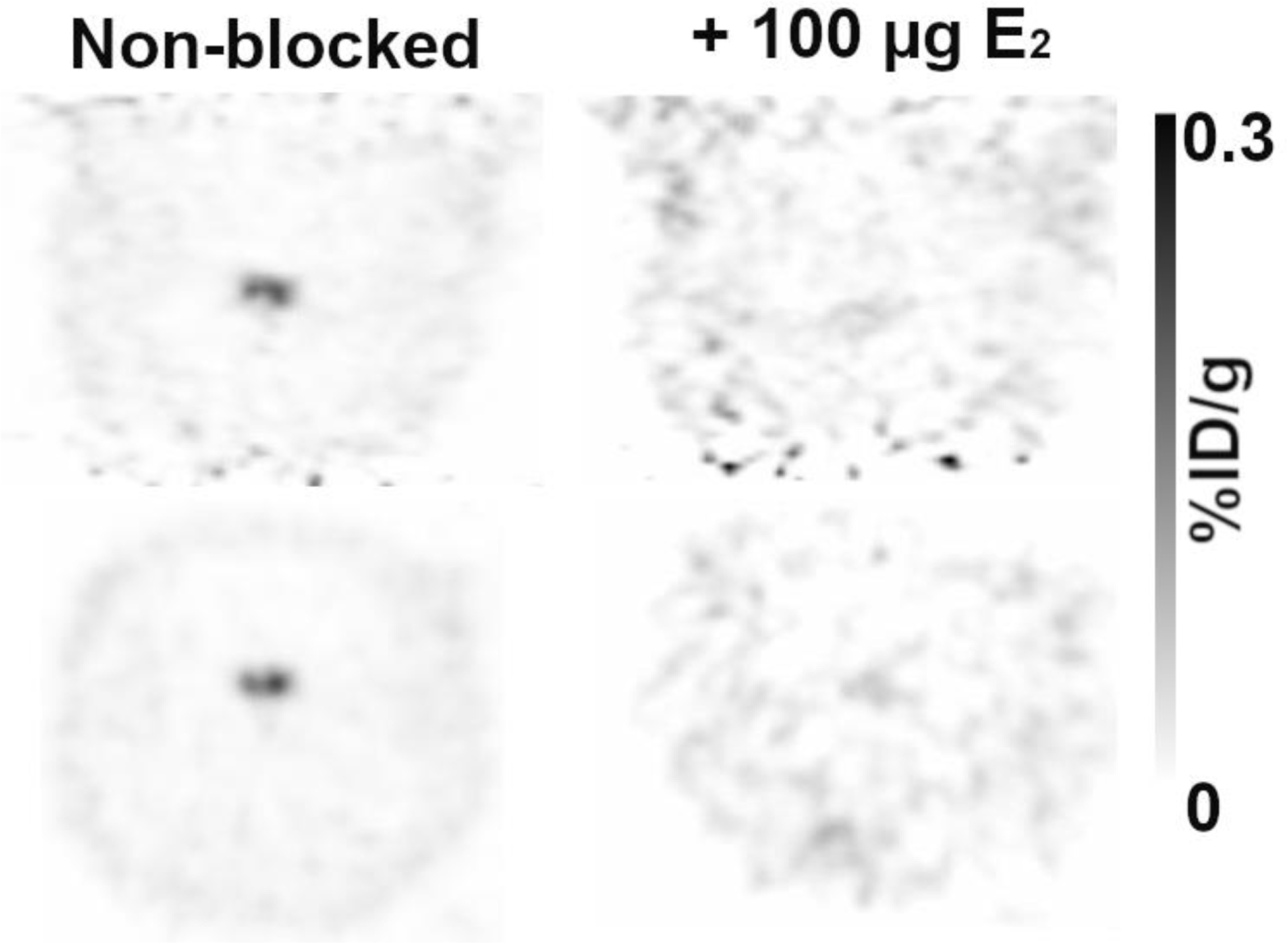
Figure 2:

Coronal (top) and transaxial (bottom) views of 4FMFES-PET images centered on the pituitary without (left) or with (right) co-injection of 100 μg estradiol (E2). Scans were performed one week apart. Co-injection of E2 with 4FMFES provoked a 25-fold decrease in signal intensity, almost down to cortical levels. Intensity scale in %ID/g in grayscale is the same for both images.
