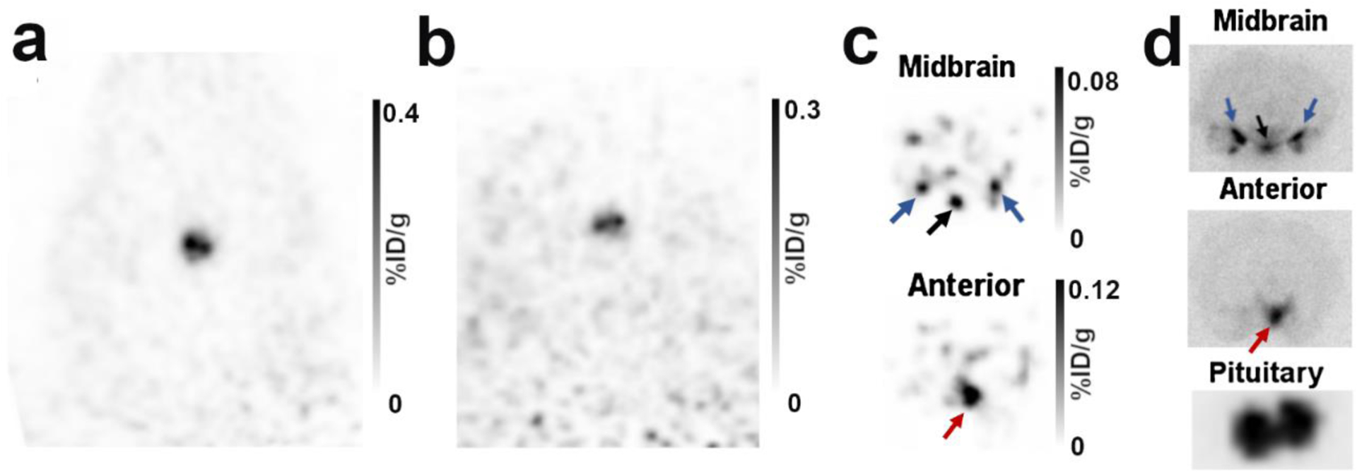
Figure 7: Dissected brain images following 4FMFES injection in female rats.

a) Typical coronal views of the pituitary of a live female rat 1 hour following injection of 4FMFES. b) After removal of the brain immediately after the in vivo PET scan, the pituitary stem remained in the skull. PET imaging of the brainless carcass reproduced the same bilateral signal as in the live animals, confirming that the signal originated from the pituitary. c) PET image of the isolated brain. A transverse midbrain slice aligned with the pituitary revealed 3 foci at the bottom of the brain which are likely to be the arcuate and ventromedial nuclei (black arrows), along with the medial and cortical amygdala (blue arrows). A transverse slice situated ~3 mm anterior to the “midbrain” slice reproduced the weak anterior signal (red arrows) seen in figure 3 and 4, which correspond to the medial preoptic area (MPOA). d) Autoradiography of 1 mm thick slices of the regions revealed in (c) and of the isolated pituitary.
