
Figure 4.
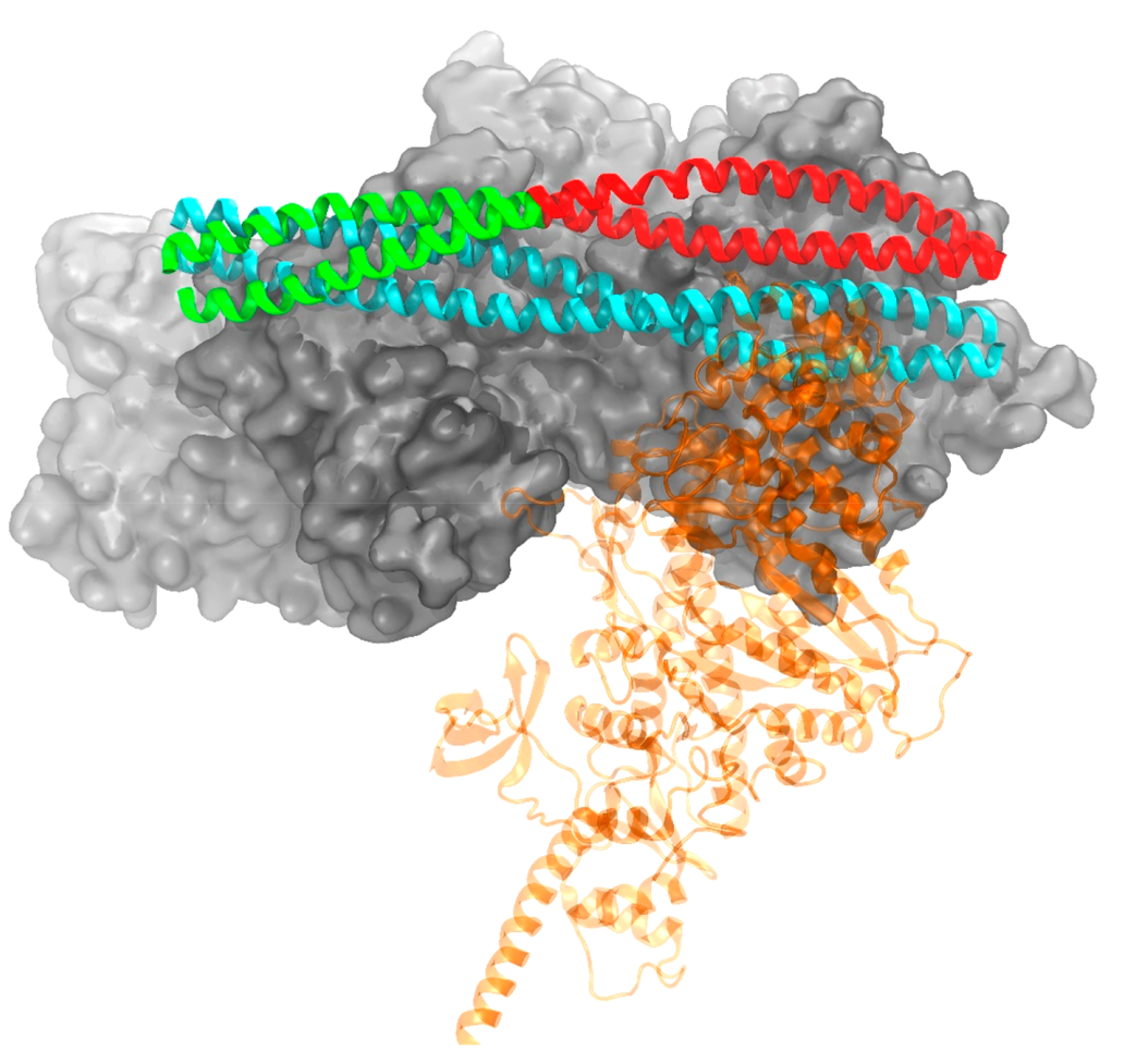
Conformations of tropomyosin in a blocked (cyan) position and at the end of a metadynamics simulation (green/red) mimicking a single myosin head (transparent for clarity) binding. The green portion of Tm illustrates the segment biased in the cooperative simulation, while red was biased in the linker simulation. Troponin not shown for clarity.