Fig. 2.
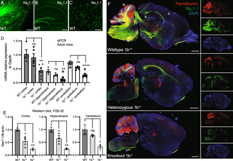
Scn1a expression is reduced in 1b deletion mouse model. a–c Immunofluorescent analysis of NaV1.1 in wildtype mice across cerebellum (a), hippocampus (b), and cortex (c), regions taken from wildtype in panel f. Scale bars a and b = 100 μm, c = 250 μm. d Bar plot showing relative expression of Scn1a using qPCR in 3-month-old mice (mean ± SEM), values normalized to WT cortex. Scn1a expression reduced in 1b−/− cortex vs WT cortex (**P = 0.0092), 1b−/− hippocampus vs WT hippocampus (**P = 0.0029), and 1b−/− cerebellum vs WT cerebellum (****P < 0.0001). e Western blots of P29-32 mouse brain membrane fractions, showing reduction of NaV1.1 protein in cortex of 1b+/− (*P = 0.0174) and 1b−/− (**P = 0.0014) mice, hippocampus of 1b+/− (*P = 0.0445) and 1b−/− (**P = 0.0025) mice and cerebellum of 1b−/− (*P = 0.0142) mice. f Immunofluorescent analysis of sagittal sections of P28 mice revealed a reduction in NaV1.1 (green) expression in homozygous versus WT mice with no changes in parvalbumin (red) expression. Scale bars = 1 mm

