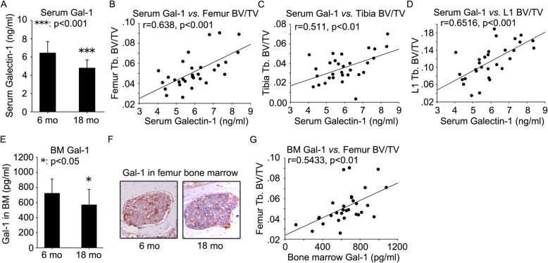
Fig. 2.
Age-related decline in Gal-1 levels in peripheral blood serum and bone marrow microenvironment in 18-month-old mice and the correlation of trabecular bone volume fraction with Gal-1 levels. Serum was prepared from peripheral blood of 6- and 18-month-old mice. ELISA was employed to quantify the levels of Gal-1 (a). The correlation of serum Gal-1 levels with BV/TV of femur (b), tibia (c), and L1 vertebrae (d) was analyzed. Femur bone marrow aspirates were prepared from 6- and 18-month-old mice. Gal-1 was quantified by ELISA (e). Immunohistochemistry assay was employed to investigate Gal-1 expression in femur bone marrow of 6- and 18-month-old mice (f). The correlation of bone marrow Gal-1 levels with BV/TV of femur (g) was analyzed. Data were shown as the means ± SD. *: p < 0.05, ***: p < 0.001, 18 mo vs. 6 mo

