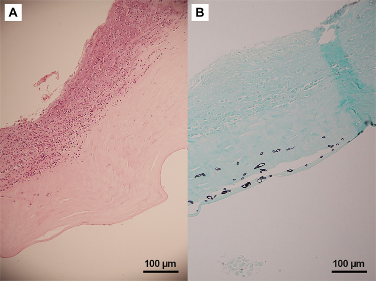
Figure 3.
Histopathology of Pythium keratitis in a globe salvage case (Case#19). (A) Histopathology section shows an ulcerated corneal lesion with numerous acute inflammatory cells and necrotic cells primarily located at the anterior stroma (Hematoxylin-Eosin); (B) special stain shows varying sizes of short hyphae at the posterior stroma and pre-Descemet’s membrane area (Gomori Methenamine Silver).

