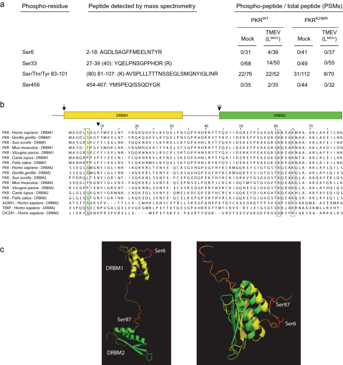
Figure 1.
PKR serine 6 can be phosphorylated in TMEV-infected cells. (a) Phosphorylation sites identified after PKR immunoprecipitation from HeLa (PKRWT) or from PKR-KO cells transduced to express a kinase-dead PKR mutant (PKRK296R), thus deficient for autophosphorylation. Phosphorylated PKR peptides were identified in mock-infected cells and in cells infected with the LM60V TMEV mutant. The PSMs (peptide spectrum match) ratio of phosphorylated on total corresponding peptides are shown for the detected phospho-peptides. (b) Amino acid sequence alignment of the first and second dsRNA binding motifs (DRBMs—yellow and green frames) from the indicated species. (c) Conserved position of Ser6 and Ser97 relative to DRBMs. The DRBM1 and 2 are shown in yellow and green, respectively. Ser6 and Ser97 are shown in red and labeled in the unstructured regions preceding the DRBMs. On the right a merged picture was generated by superimposing the two DRBMs and the surrounding regions (NMR structure of PKR8, PDB code 1QU6 ). Images were generated using the PyMOL molecular Graphic system, Version 2.1, Schrödinger, LLC.

