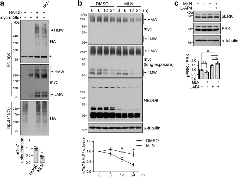
Fig. 4. Effects of neddylation on mGlu7 ubiquitination, degradation, and signaling.
a Neddylation facilitates mGlu7 ubiquitination. HEK 293 T cells were cotransfected with myc-mGlu7 and/or HA-Ub. Thirty-six hours after transfection, the cells were treated with 1 μM MLN4924 for 6 h and 1 mM L-glutamate for 5 min in the presence of MG132 and leupeptin. The cell lysates were immunoprecipitated with an anti-myc antibody. Western blotting was performed with the indicated antibodies. *, nonspecific bands; Ub, ubiquitin. The bar graph with scatter plots shows the mean ± SEM (DMSO, 1.00 ± 0.08; MLN, 0.43 ± 0.06; n = 3, *p < 0.05, Student’s t test). b Time-course experiment of the effect of neddylation on mGlu7 stability. HEK 293 T cells were transfected with myc-mGlu7 and HA-NEDD8 and pretreated with 1 μM MLN4924 for 12 h. The culture medium was changed to DMEM with 1% fetal bovine serum, and CHX was added for the indicated time periods; h, hours. Time-course expression of mGlu7 was quantified and is presented as the mean ± SEM normalized to 0 h (DMSO, 6 h: 1.10 ± 0.06, 12 h: 0.96 ± 0.07, 24 h: 0.83 ± 0.10; MLN, 6 h: 0.96 ± 0.09, 12 h: 0.66 ± 0.09, 24 h: 0.40 ± 0.08; n = 3, *p < 0.05, Student’s t test). c Neddylation is not required for mGlu7-mediated ERK signaling. Primary cortical neurons (DIV14) were treated with 1 μM MLN4924 and/or 400 μM L-AP4. Western blotting was performed with the indicated antibodies. The bar graph with scatter plots shows the mean ± SEM (vehicle, 1.00 ± 0.01; MLN, 0.73 ± 0.13; L-AP4, 1.45 ± 0.06; MLN + L-AP4, 1.72 ± 0.07; n = 3; *p < 0.05, n.s. indicates p > 0.05, one-way ANOVA followed by Tukey’s post hoc test).

