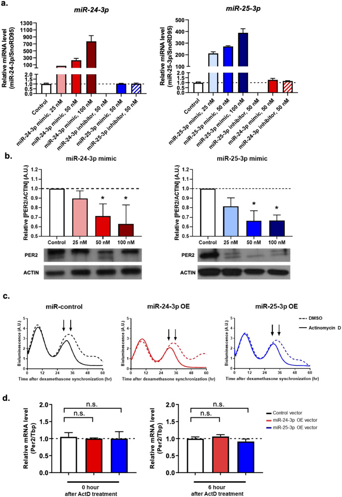
Fig. 2. miR-24-3p and miR-25-3p modulate Per2 gene expression at the posttranscriptional level.
Wild-type mouse embryonic fibroblasts (WT MEFs) were transfected with miR-24-3p or miR-25-3p mimics in a dose-dependent manner (25, 50, and 100 nM) and then assayed for the expression levels of a miR-24-3p and miR-25-3p using real-time qPCR (n = 4) and b quantified PER2 protein expression by Western blotting (n = 3). To determine the effects of miR-24-3p and miR-25-3p on Per2 transcription, Period2::Luc knock-in mouse embryonic fibroblasts (Per2::Luc KI MEFs) were transfected with 0.5 µg of miR-24-3p- and miR-25-3p-overexpressing and control vectors, and then, actinomycin D (a potent transcription inhibitor) was added for 30 h after dexamethasone synchronization. c The results of real-time bioluminescence recordings are represented in raw data format after actinomycin D (solid line) and DMSO (dashed line) treatment. d Per2 mRNA was quantified after an additional 0 and 6 h of actinomycin D treatment (Per2::Luc KI MEFS were harvested as indicated by the arrowheads), and the data were normalized to that of the control vector (n = 4). Data are presented as the means ± SE, and the significance was assessed by one-way ANOVA, *p < 0.05 compared to the control groups.

