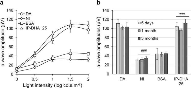
Fig. 5. Effect of IP-DHA on light-induced degeneration over time.
a Full-field ERG responses of Abca4−/− mice 3 months after a single IP-DHA injection. ERG scotopic responses were recorded, and a-wave amplitudes were plotted as a function of light intensity. IP-DHA (25 mg/kg)-treated mice exposed to light showed an a-wave amplitude similar to that of DA control mice, whereas BSA-treated and untreated (NI) mice exposed to light did not recover amplitudes. The data are presented as the mean ± SEM from n = 5–6 mice. b a-wave amplitudes recorded at 2-log cd.s.m−2 did not change in each treatment over the 3-month period. The data are presented as the mean ± SEM from n = 5 mice. One-way ANOVA with Bonferroni’s multiple comparison posttest: ###p < 0.001, NI vs. DA; ***p < 0.001, IP-DHA25 mg/kg vs. BSA; IP-DHA vs. DA, nonsignificant.

