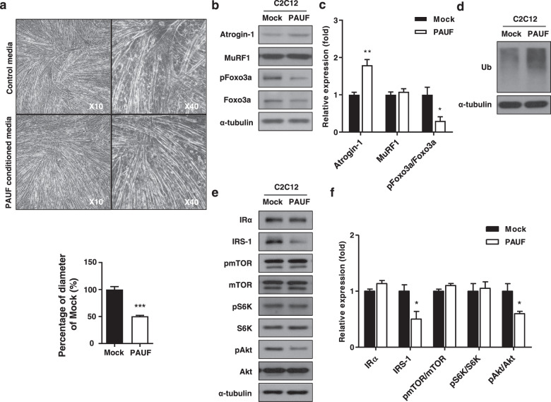
Fig. 5. Myotube atrophy induced by PAUF conditioned medium.
a Light microscopy images (left: low magnification at 10×; right: high magnification at 40×) showing the effect of control medium (from Panc-1/Mock cell cultures) or PAUF conditioned medium (from Panc-1/PAUF cell cultures). Relative diameter of myotubes cultured in control or PAUF conditioned medium. Four different views were randomly selected for diameter measurements and quantified using ImageJ software. Data were normalized to the diameter of myotubes cultured in control medium. b Western blot analysis of catabolic markers in cells treated with rPAUF using antibodies against Atrgoin-1, MuRF1, pFoxo3a, and Foxo3a. c Relative expression was measured by densitometric analysis of western blot data (n = 3). The data represent the mean ± SD; *p < 0.05 and **p < 0.01. d Whole-cell extracts were also subjected to SDS-PAGE followed by western blot analysis using an anti-ubiquitin antibody to assay steady-state ubiquitination levels. e Western blot analysis of anabolic markers in cells treated with rPAUF using antibodies against IR, IRS-1, pmTOR, mTOR, pS6K, S6K, pAkt, and aAkt. f Relative expression was measured by densitometric analysis of western blot data (n = 3). The data represent the mean ± SD; *p < 0.05.

