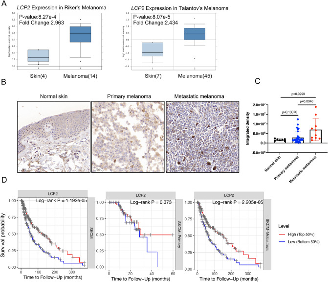
Figure 2.
The expression and prognostic value of LCP2 in melanoma. (A) LCP2 expression level in human normal skin tissue and skin cutaneous melanoma. Based on the Oncomine database, box-plot diagrams were displayed to compare the LCP2 level in human normal skin tissue with that in skin cutaneous melanoma from studies reported by Riker et al. (p value: 8.27e−4, Fold Change: 2.963) and Talantov al. (p value: 8.07e−5, Fold Change: 2.434). (B) LCP2 protein expression in normal skin, primary melanoma and metastatic melanoma tissue. (C) Quantitative analysis of immunohistochemistry indicated that LCP2 protein was highly expressed in metastatic melanoma tissue compared with normal skin and primary melanoma. (D) Based on differential LCP2 expression level, patients with high-LCP2 expression had a better prognosis as compared with those with low-LCP2 expression in skin cutaneous melanoma and in cutaneous melanoma with metastasis, but not in primary sites of skin cutaneous melanoma.

