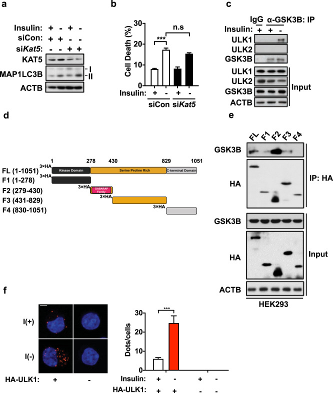
Fig. 2. ULK1 interacts with GSK3B.
a Western blotting analysis of the effect of KAT5 knockdown on MAP1LC3B lipidation after insulin withdrawal for 6 h. b Cell death rate following insulin withdrawal for 24 h in HCN cells transfected with control (siCon) or KAT5-targeting (siKAT5) siRNA (n = 3). c The interaction of endogenous ULK1 or ULK2 with GSK3B was analyzed by immunoprecipitation with anti-GSK3B antibody after insulin withdrawal for 6 h. d Schematic representation of the ULK1 domain structure and fragment constructs. e Analysis of the ULK1 regions responsible for GSK3B interaction. The indicated HA-ULK1 truncation mutants were expressed in HEK293 cells and precipitated with an anti-HA antibody, and coimmunoprecipitation with endogenous GSK3B was determined by western blotting. The blots shown are representative of at least three experiments with similar results. f HCN cells were fixed and analyzed by the proximity ligation assay with primary antibodies against GSK3B and the HA-tag. The cells were imaged by confocal microscopy. Scale bar, 10 μm. The graph represents the number of dots per cell following insulin withdrawal (n = 9 cells). ***P < 0.001. ns not significant.

