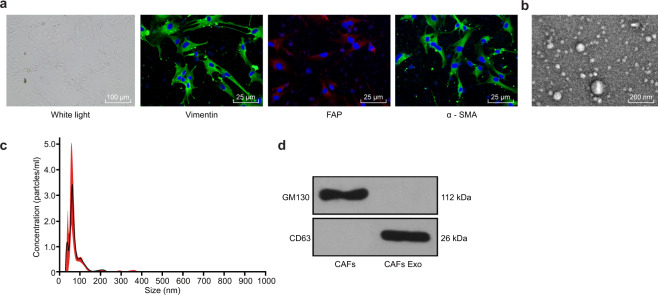
Fig. 1. Characteristics of cancer-associated fibroblasts and isolation of exosomes.
a Representative morphology of CAFs (scale bar, 200 μm) and immunofluorescence staining identification of CAFs using antibodies against Vimentin, α-SMA, and FAP (scale bar, 100 μm). b Transmission electron microscopy showing exosomes isolated from the CAFs (scale bar, 100 nm). c Nanoparticle tracking analysis of the CAF-derived exosomes (represented as size vs. concentration). d Western blot analysis of the exosome marker CD63 and the Golgi matrix protein GM130 in exosome-enriched conditioned medium.

