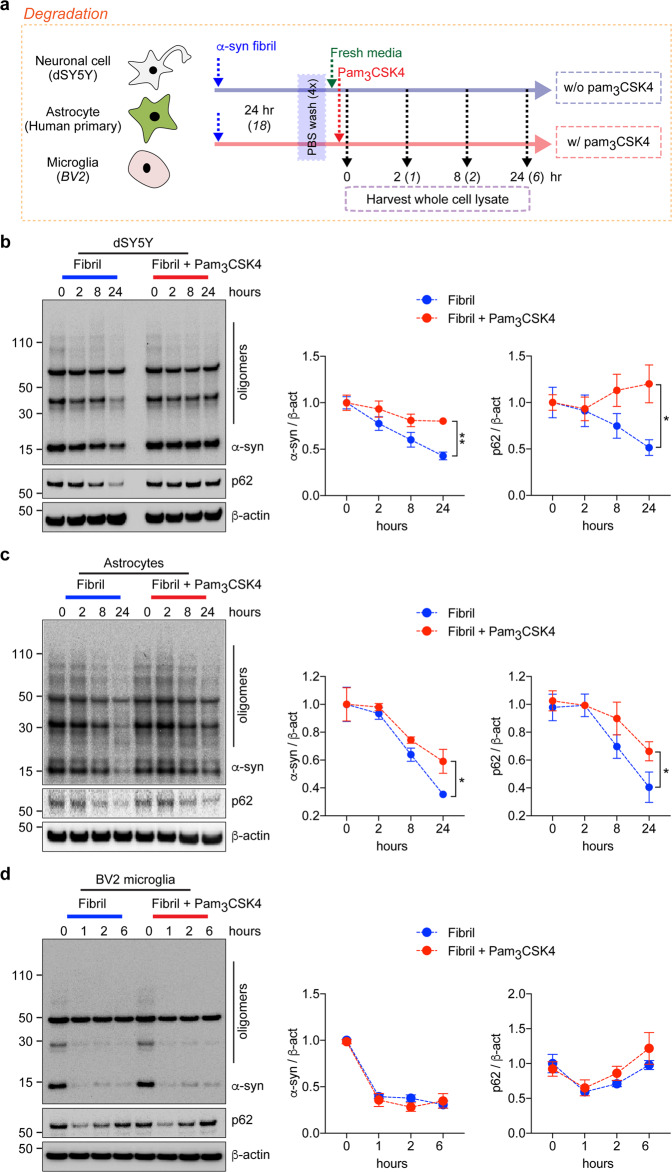
Fig. 2. Stimulation of TLR2 and kinetics of α-synuclein fibril degradation by neurons, astrocytes, and microglia.
a Experimental scheme of the α-syn degradation assay. b–d dSY5Y cells (b), human primary astrocytes (c), and BV2 microglia (d) were pretreated with α-syn fibrils (200 nM) for the indicated duration. After washing with PBS, the cells were incubated in the presence or absence of pam3CSK4 (10 μg/ml) and harvested at the indicated time points. Whole-cell lysates were probed for α-synuclein, p62, and β-actin. The levels of α-synuclein and p62 were determined by densitometric quantification. The data are the mean ± SEM. *p < 0.05 and **p < 0.01.

