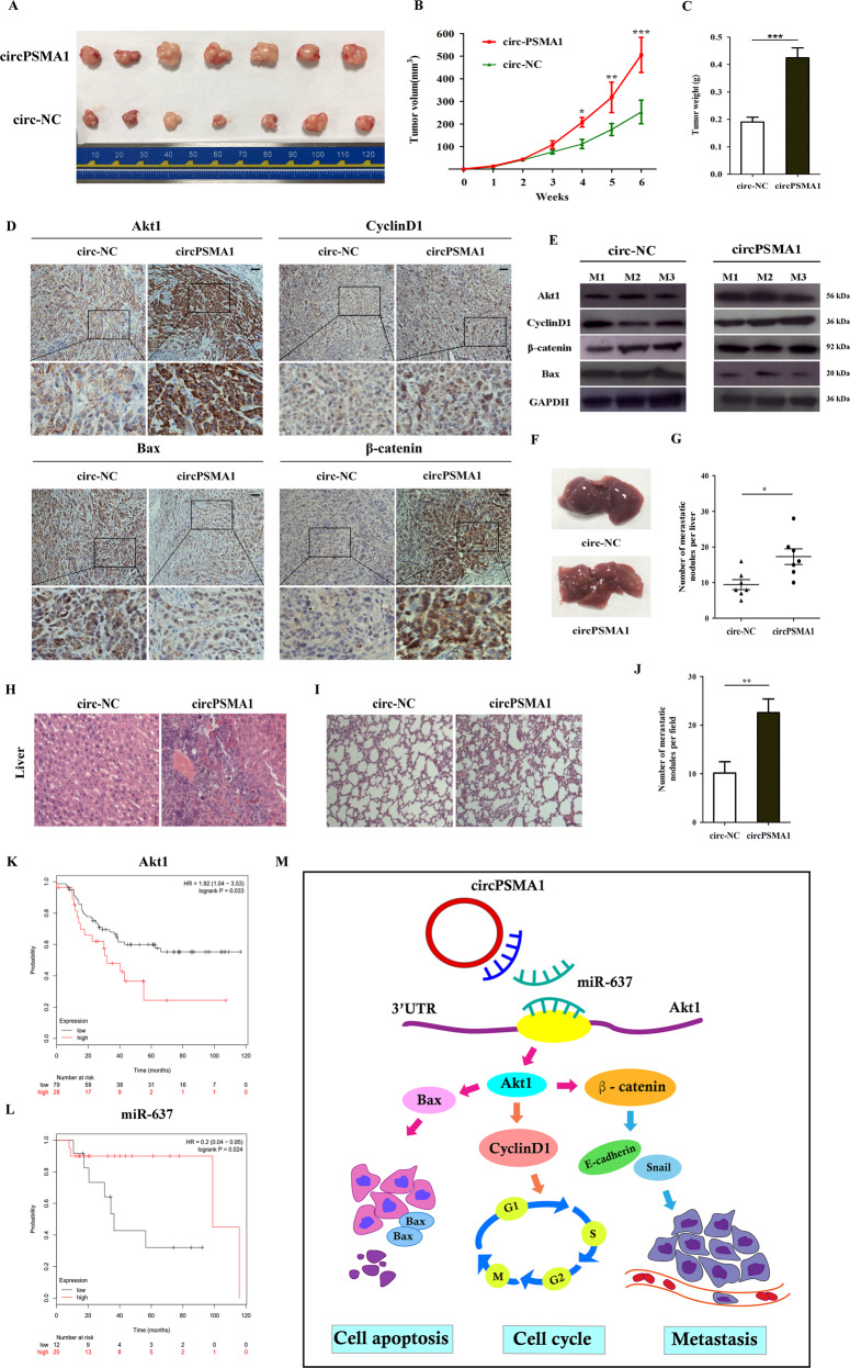
Fig. 8. CircPSMA1 facilitates tumorigenesis, migration, and metastasis of TNBC cells in vivo.
A Representative images of tumors of circPSMA1-overexpressing group (n = 7) or NC groups (n = 7). B Growth curves of tumors were measured weekly. C Tumor weight. D Immunohistochemistry (IHC) staining showed the protein levels of Akt1 and Akt1-related molecules (β-catenin, cyclin D1, and Bax) (magnification, ×200, Scale bar, 100 μm). E The protein level of Akt1 and Akt1-related molecules were detected by western blot. F–H The appearance and HE stain images of livers in two groups (magnification, ×100, Scale bar, 100 μm). I–J The HE stain images of lungs in two groups (magnification, ×50, Scale bar, 100 μm). K–L Kaplan–Meier survival analysis of overall survival (OS) in TNBC patients with lymphatic metastases based on TCGA data according to the Akt1 (n = 107) or miR-637 expression (n = 32). M The illustration of how circPSMA1/miR-637/Akt1 axis promoted TNBC cell proliferation, migration, and metastasis. CircPSMA1 functions as a sponge of miR-637 and downregulates miR-637 expression, and then increases Akt1 expression by directly binding miR-637 with the 3′-UTR of Akt1. Akt1 can upregulate cyclin D1 expression and lead to G1/S phase cell cycle arrest; Akt1 can upregulate β-catenin expression and cause to tumor migration and metastasis; Akt1 can downregulate Bax expression and thereby inhibiting cell apoptosis. Therefore, circPSMA1 promoted TNBC cell proliferation, migration and metastasis through circPSMA1/miR-637/Akt1/β-catenin (cyclin D1) pathway.

