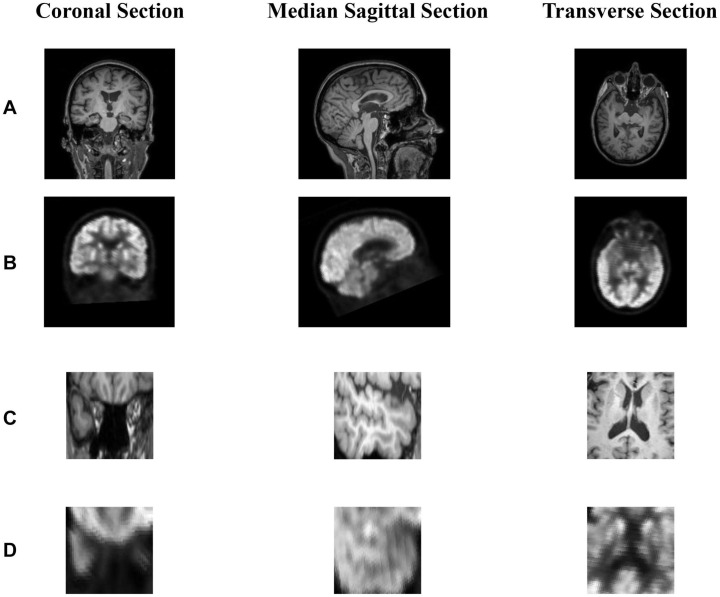
FIGURE 3.
(A,B) show the coronal section, median sagittal section and transverse section of MR image and PET image, respectively. (B) is registered to the corresponding (A) through rigid registration. The dimensions of (A,B) are the same as 221 × 257 × 221. (C) is the area near the hippocampus of the MR image and (C) is obtained from (A). Since (A,B) are registered, we can obtain (D) corresponding to (C) from (B). In short, (A,B) or (C,D) represents the same area in the brain.

