
FIGURE 1.
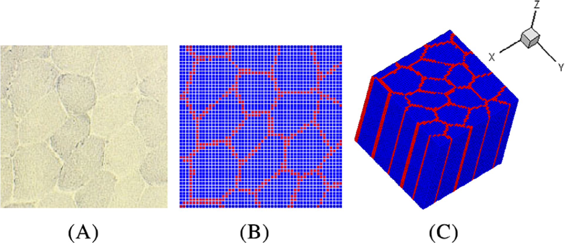
The procedure of constructing the 3D model from the 2D images of the cellular-scale cross-section of skeletal muscle tissue.12 (A) the cross-sectional image of a representative unit cell of the muscle tissue, (B) level-set segmented cross-section, and (C) 3D model produced by extruding the 2D model along the z-direction. Blue points denote the muscle fiber phase and red points denote the connective tissue phase