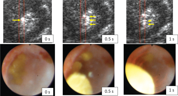
FIG. 5.
Ultrasonic propulsion of the stone fragments after BWL, visualized with the US Propulse 1 system (top) and by ureteroscopy (lower). The times (0, 0.5, and 1 second) show frames just before, in the middle of, and at the end of the 1-second ultrasonic propulsion pulse, which is traveling down in the US frames and out of the page in the ureteroscope frames. The red x and red lines in the US frames indicate the focus and focal region on the Propulse 1 display, and yellow arrows were added in postprocessing to show the one collection of fragments at 0 second spreading and moving downward in the frames at 0.5 and 1 second. This motion and US imaging revealed to the operator that the stone was no longer intact stone but was instead many fragments as has been observed in vitro.1 One ultrasonic propulsion pulse was observed to move the fragments out of the calix, and in ultimate clinical use, many pulses may move the stones out of the collecting system to facilitate clearance. (Supplementary Videos S1 and S2) are included.

