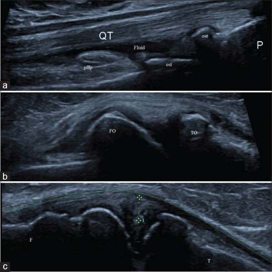
Figure 3.

High-frequency ultrasonographic evaluation of osteoarthritis-affected knee joint showing; (a) presence of minimal effusion (fluid) in the supra-patellar recess and multiple osteophytes (ost) at patello-femoral joint; (b) presence of femoral and tibial osteophytes at joint margins (FO and TO); and (c) evidence of medial meniscal extrusion (calipers) causing displacement of medial collateral ligament (green line). F: Femur, T: Tibia, QT: Quadriceps tendon, pffp: Prefemoral fat pad and P: Patella
