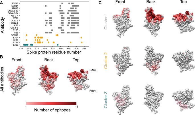
Fig 1. Epitopes for antibodies targeting the spike protein RBD overlap substantially.
A. Contact residues for spike protein RBD antibody epitopes. Colors and symbols denote antibody clusters: Grey squares: Cluster 1, yellow diamonds: Cluster 2, green circles: Cluster 3. B. RBD structure with each residue colored by the number of antibody epitopes including it, compiled from PDB data. C. RBD structure, colored by the number of antibody epitopes that each residue is part of, by epitope cluster.

