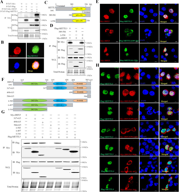
Fig 2. DDX5 directly interacts with METTL3.
A: Co-IP of DDX5 and METTL3 in HEK293T cells. B: Co-localization of DDX5 and METTL3 in HEK293T cells cultured in slices and co-transfected with Myc-DDX5 and Flag-METTL3 plasmids. After 24 h, slices were fixed, permeabilized, and incubated with rabbit anti-Myc and mouse anti-Flag antibodies followed by Alexa Fluor 546 labeled goat anti-rabbit IgG (H+L), Alexa Fluor 488 labeled goat anti-mouse IgG (H+L). Nuclei were stained with DAPI; cells were observed by LSCM. Scale bars, 5 μm. C: Schematic of full-length METTL3 and its truncated mutants. D: Co-IP of DDX5 and METTL3 mutants in HEK293T cells. Cells were co-transfected with Myc-DDX5 and Flag-METTL3 mutants. After 24 h, cells were lysed and subjected toIP with Flag antibody, and whole-cell lysates and IP were analyzed by western blotting. E: Co-localization of DDX5 and METTL3 mutants in HEK293T cells. Scale bars, 10 μm. F: Schematic of full-length DDX5 and its truncated mutants. G: Co-IP of METTL3 and DDX5 mutants in HEK293T cells co-transfected with Flag-METTL3 and Myc-DDX5 mutants. After 24 h, cells were lysed and underwent IP with Myc antibody. Whole-cell lysates and IP were analyzed by western blotting. H: Co-localization of METTL3 and DDX5 mutants in HEK293T cells cultured and co-transfected with Flag-METTL3 and Myc-DDX5 mutant plasmids. After 24 h, slices underwent IFA. Scale bars, 10 μm.

