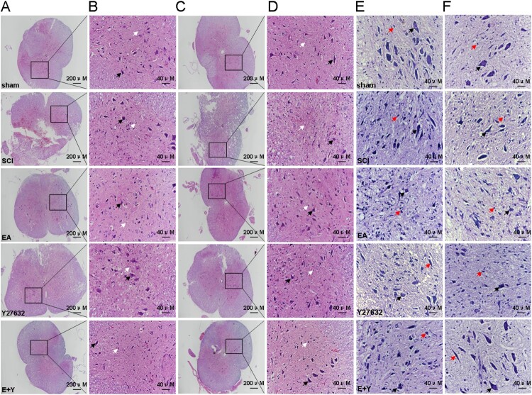
Figure 6.
HE and Nissl staining in each group. Neuron cell was indicated by the black arrows, and neuroglia cell was indicated by the white arrows in HE staining and by the red arrows in Nissl staining. (A,B) HE staining at 7 days; (C,D): HE staining at 14 days; (E) Nissl staining at 7 days; (F) Nissl staining at 14 days. (n = 4 rats/group) sham: sham operation; SCI: SCI; EA: electroacupuncture; Y: blocking agent Y27632.

