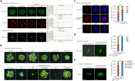
Fig. 2. RH6/RH8/RH12 is required for D-body formation.

(A) Representative images and fluorescence recovery curves of D-body components. Leaf epidermal cells of N. benthamiana (inoculated with DCL1-YFP/RFP-SE for 72 hours) and root tip cells (elongation region) of 1-week-old A. thaliana (pHYL1::HYL1-YFP) plants were analyzed. The region of interest is highlighted with a red circle. Scale bars, 2 μm. The intensity was normalized against the average prebleach fluorescence. n = 3 for each group. The green line indicates the half-recovery time. (B) Whole-plant images of 6-week-old plants. Scale bars, 0.5 cm. (C) Detection of DCL1 subcellular localization with anti-DCL1 antibodies. Left: One set of representative images. Right: The quantification analysis. The numbers 0 to 3 represent the number of DCL1 granules in each nucleus. In total, 150 to 200 nuclei were analyzed for each genotype. Scale bars, 1 μm. (D and E) Subcellular localization of HYL-YFP (D) and YFP-SE (E) in 3-week-old A. thaliana plants. Fifteen root tip cells (elongation region) for HYL-YFP and 115 to 125 leaf epidermal cells for YFP-SE were analyzed for each genotype. Scale bars, 5 μm. Photo credit: Ningkun Liu, Institute of Zoology, Chinese Academy of Sciences.
