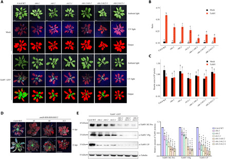
Fig. 6. RH6/RH8/RH12 facilitates viral proliferation.

(A) Visualization of rosette leaves of plants inoculated with TuMV::GFP or buffer (Mock). Images were taken at 14 dpi. In the pseudocolor images, green color refers to infected plant pixels, while red color refers to noninfected plant pixels. Scale bars, 0.5 cm. (B) Bar plot showing the ratio of infected area to the total area of Col-0 WT and mutant plants at 14 dpi. (C) Bar plot showing the rosette size at 14 dpi. Statistical analysis was performed between treatments in each genotype. The boxes with different letters are significantly different (n = 8, Tukey post hoc test, with α = 0.05). (D) TuMV-infected amiR-RH6/RH8/RH12 plants. Four-week-old A. thaliana plants with TuMV::GFP were examined, and the plants were imaged under ultraviolet (UV) light at 14 and 22 dpi. Scale bars, 0.5 cm. (E) Western blot analysis of the accumulation of CP, VPg, and HC-Pro proteins. Representative images and quantitative analysis are shown on the left and right, respectively. Photo credit: Ningkun Liu, Institute of Zoology, Chinese Academy of Sciences.
