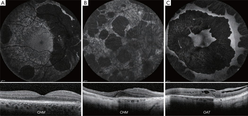
Figure 7.
Retinal Imaging of Chorioretinal Dystrophies. (A-C) Fundus autofluorescence (FAF) imaging with corresponding horizontal trans-foveal optical coherence tomography (OCT). (A) Choroideremia (CHM) in a 35-year-old male with large areas of atrophy on FAF, not yet involving foveal center on OCT. (B) Choroideremia (CHM) in a severely affected 75 year old female carrier with large areas of atrophy on FAF and involving the foveal center on OCT, with a small area of spared ellipsoid zone. (C) Gyrate Atrophy (OAT); large well-demarcated coalesced areas of atrophy visible on FAF as decreased AF, and OCT with macular oedema and retinal tubulations.

