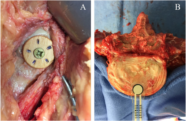
Fig. 4.
Simulated cam-type femoroacetabular impingement. (A) Anterior view of the right hip of a cadaveric specimen demonstrating simulated cam-type femoroacetabular impingement, utilizing a wooden knob placed at the femoral head-neck transition. (B) Piezoresistive force sensors placed in the antero-medial section of a lumbar intervertebral disk.

