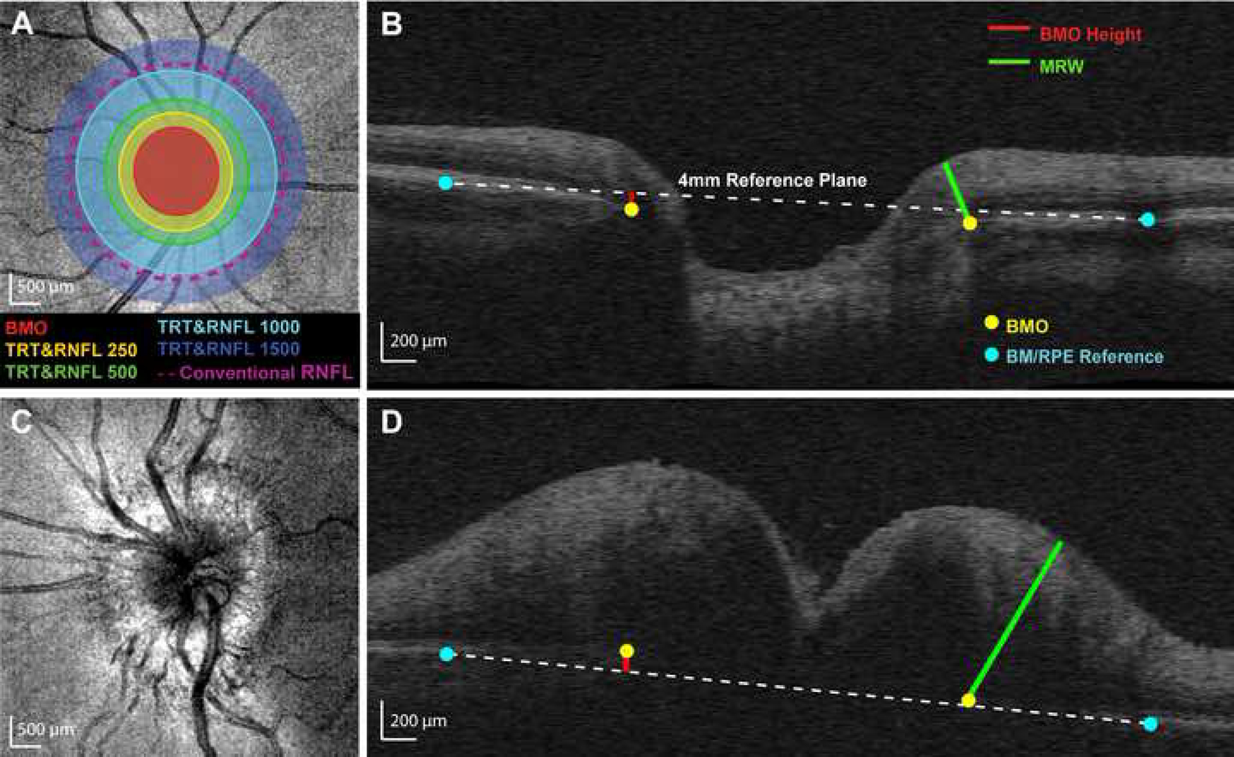
Figure 1.

Calculation of custom parameters. (A) Cirrus optical coherence tomography mean reflectance image of a control eye. Selected Bruch’s membrane opening (BMO) points were fit with an ellipse. Five annular zones were used for determining average retinal nerve fiber layer (RNFL) thickness and total retinal thickness (TRT) from volumetric data: (1) within the BMO ellipse (BMO, red), (2) BMO ellipse to 250 µm (RNFL250 and TRT250, yellow), (3) 250 to 500 µm (RNFL500 and TRT500, green), (4) 500 to 1000 µm (RNFL1000 and TRT1000, light blue), and (5) 1000 to 1500 µm (RNFL1500 and TRT1500, dark blue). The conventional RNFL scan (nominally 1730 µm from the center of the optic nerve head, approximately 1100 µm from the BMO in this individual) is represented by the dashed purple line. (B) Control B-scan demonstrating custom optic nerve head (ONH) parameters. Minimum rim width (MRW, green line) was calculated as the minimum distance from BMO (yellow dots) to the internal limiting membrane. A reference plane (white dashed line) was created by connecting points selected at the Bruch’s membrane/retinal pigment epithelium interface 2 mm from the ONH center (blue dots) for each interpolated radial scan. BMO height (red line) was calculated as the perpendicular distance from the reference plane to the BMO. (C) Mean reflectance image of an eye with papilledema. (D) B-scan for the same papilledema eye, demonstrating custom ONH parameters. Note that the BMO is above the reference plane in this subject with papilledema.
