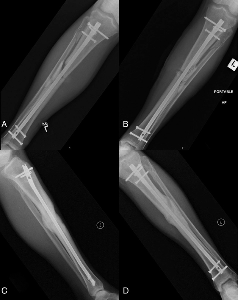
Figure 1.

Acute FRI. Radiographs of a 39-year-old male who presented with a grade II open left diaphyseal tibia fracture following a motor vehicle accident. He was initially treated with irrigation and debridement, intramedullary nailing, and primary skin closure. He presented 4 weeks postoperatively with increasing pain at the fracture site combined with erythema and wound drainage. Radiographs at that time demonstrated stable hardware (A). He was taken back to the operating room for irrigation and debridement, examination of the hardware for stability, deep tissue cultures, and the placement of local antibiotics including vancomycin powder and antibiotic calcium sulfate beads (Osteoset T, Wright Medical) (B). His original hardware was maintained, and his intraoperative cultures grew Enterococcus Faecalis. He was placed on tailored IV antibiotic therapy for 6 weeks followed by 3 months of oral antibiotics. No further surgical treatment was required and his 1 year follow-up radiographs demonstrate solid union (C and D).
