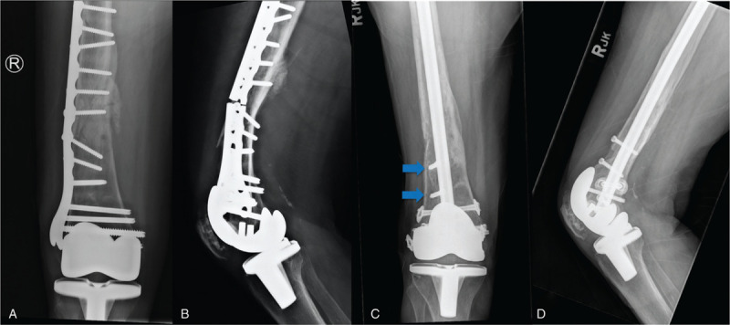
Figure 7.

Periprosthetic distal femoral nonunion. Radiographs of 68-year-old female 9 months after open reduction and internal fixation of a periprosthetic distal femur fracture, demonstrating nonunion and plate breakage (A and B). The initial construct was overly rigid with too short of a plate, insufficient working length, excessive screw fill, and inadequate spacing of fixation. Postoperative radiographs 6 months after revision fixation using a retrograde intramedullary nail and bone morphogenetic protein (BMP, Infuse, Medtronic) (C and D). Note the use of blocking screws to restore anatomic alignment (blue arrows).
