. 2021 Apr 13;10:e67817. doi: 10.7554/eLife.67817

Figure 4. DolP associates with the BAM complex via an interaction with BamA.

(A) The envelope fractions of BW25113 cells carrying the indicated plasmids were solubilized with 1% (w/v) digitonin and 0.1% (w/v) DDM and subjected to IgG affinity purification of protein A-tagged DolP. The load and elution fractions were analysed by SDS-PAGE. The coomassie staining of the elution of protein A-tagged DolP is shown below the diagrams representing the overproduced protein. Blotted proteins from load and elution fractions were detected by immunolabelling using the indicated antisera. Load 0.5%; Elution 100%. The asterisk indicates the TEV-digestion product of DolPProtA. (B) The envelope fractions of BW25113 cells carrying the plasmids overproducing His-tagged DolP and the indicated BamA protein variants (deleted of POTRA1 or of POTRA2) were solubilized with 1% (w/v) digitonin and 0.1% (w/v) DDM and subjected to Ni-affinity purification. The load and elution fractions were analysed by SDS-PAGE. The coomassie staining of the elution of His-tagged DolP overproduced together with wild-type BamA is shown below the diagram representing the overproduced proteins. Blotted protein from load and elution fractions were detected by immunolabelling using the indicated antisera. Load 2%; Elution 100%. The amount of BamA co-isolated with DolPHis was normalized to the amount of BamA detected in the load fraction. The value obtained for the pBamA-DolPHis sample was set to 100%. The average of the relative amounts of co-isolated BamAΔP1 and BamAΔP2 are as follows: BamAΔP1, 16.5% (N = 2; 1st exp. 23.6%; 2nd exp. 9.3%); BamAΔP2, 81.2% (N = 2; 1st exp. 101.8%; 2nd exp. 60.6%). (C) UV photo-crosslinking of ΔdolP cells transformed with pEVOL-pBpF and pBamA-DolPHis harbouring an amber codon at the indicated position of the dolP ORF. Upon Ni-affinity chromatography of DolPHis, eluates obtained from UV irradiated samples were separated by SDS-PAGE and analysed by immunoblotting using the indicated antisera. The total envelope fraction of cells expressing DolPHis with Bpa at position V52 (non-irradiated) is shown in the first lane and serves as a reference for the migration of non-crosslinked DolP and BamA. Arrowheads indicate crosslinked products detected with both DolP and BamA antisera. Analysis of eluates obtained from non-irradiated samples are shown in Figure 4—figure supplement 1B. The amino acid residues replaced with Bpa are indicated on the structure of DolP, PDB: 7A2D (Bryant et al., 2020). In purple are the positions crosslinked to BamA. (D) The envelope fraction of BW25113 cells overproducing DolPHis or the BAM complex containing C-terminally His-tagged BamE was subjected to protein extraction with 1% (w/v) DDM, Ni-affinity purification, and gel filtration chromatography. The elution fractions were analysed by SDS-PAGE and coomassie staining. The double asterisk indicates a contaminant protein in the elution of DolP. (E) Roughly equimolar quantities of purified His-tagged BAM complex and DolP were incubated alone for 1 hr at 4°C (lanes 1, 2, and 7), or together for 1 hr at 4°C (lanes 3 and 6) or for 30 min at 25°C (lanes 4 and 5), prior to blue native-PAGE and immunoblotting using the indicated antisera.

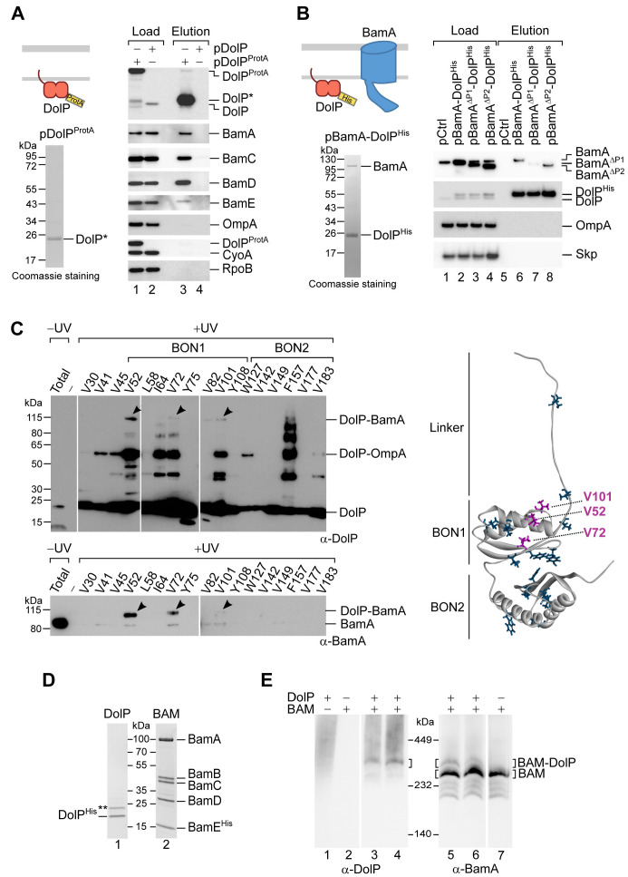
Figure 4.

Figure 4—figure supplement 1. Analysis of the DolP–BamA interaction.

Figure 4—figure supplement 1.

(A). The envelope fractions of BW25113 cells carrying pBAMProtA were solubilized with 1% (w/v) digitonin and 0.1% (w/v) DDM and subjected to IgG affinity purification of protein A-tagged BamE. The load and elution fractions were analysed by SDS-PAGE. The coomassie staining of the elution of protein A-tagged BamE is shown below the diagrams representing the overproduced proteins. Blotted protein from load and elution fractions were detected by immunolabelling using the indicated antisera. Load 1%; Elution 100%. The asterisk indicates the TEV-digestion product of BamEProtA. (B) UV photo-crosslinking of ΔdolP cells transformed with pEVOL-pBpF and pBamA-DolPHis harbouring an amber codon at the indicated position of the dolP ORF (see also Figure 4C). Eluates of the DolPHis Ni-affinity chromatography obtained from samples protected from UV irradiation were separated by SDS-PAGE and analysed by immunoblotting using the indicated antisera. The total envelope fraction of cells expressing DolPHis with Bpa at position V52 (non-irradiated) is also shown. (C) Equal aliquots of the envelope fraction of BW25113 cells expressing His-tagged DolP were solubilized with the indicated concentrations (w/v) of digitonin and DDM: 1%, 0.1% (lane 1), 0.8%, 0.3% (lane 2), 0.3%, 0.8% (lane 3), 0.1%, 1% (lane 4). His-tagged DolP was purified by Ni-affinity chromatography. In all cases, proteins were eluted in the presence of 0.3% (w/v) digitonin and 0.03% (w/v) DDM. Load 0.2%; Elution 100%. (D) The indicated strains were streaked onto LB agar plates containing vancomycin as indicated.
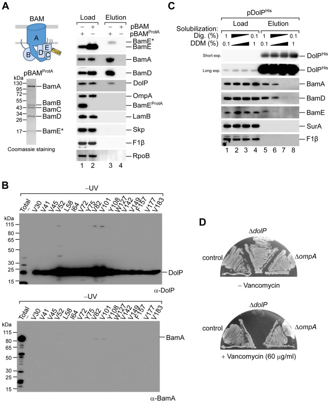
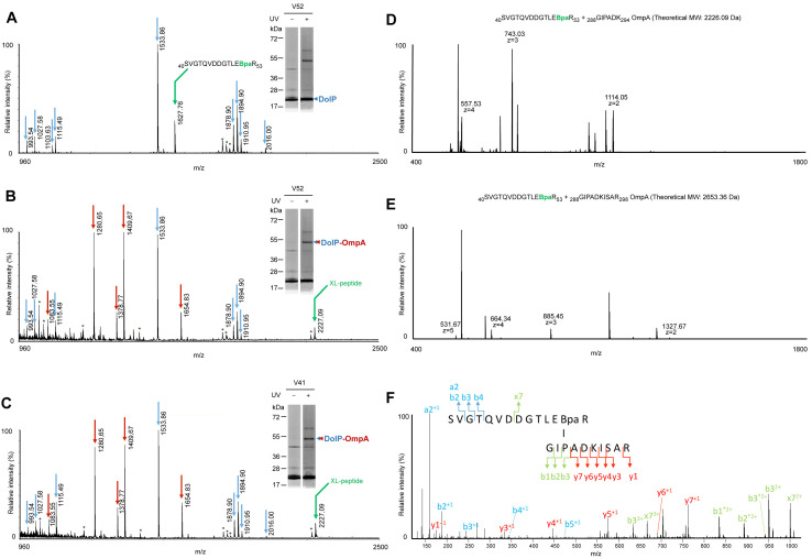
Figure 4—figure supplement 2. Mass spectrometry analyses of the DolP–OmpA crosslink product.

Figure 4—figure supplement 2.

(A–C) BW25113 cells carrying pEVOL-pBpF and a pDolPHis with amber codons engineered at positions V52 (A and B) or V41 (C) of the dolP ORF were subjected to UV crosslinking. After Ni-affinity purifications, eluates were subjected to SDS-PAGE and coomassie-staining. Bands corresponding to DolPHis and its major UV-specific crosslink products (respectively, single and double arrowheads) were trypsin digested and subjected to MALDI-TOF MS analyses. DolP and DolP together with OmpA were identified by peptide mass fingerprinting using tryptic peptide predicted patterns for each protein (blue arrows for DolP and red arrows for OmpA) in the samples obtained from the single arrowhead (A) and double arrowhead bands (B and C), respectively. (A) The mass of a tryptic peptide containing Bpa in place of V52 (m/z = 1627.76) was identified in the band containing only DolPHis. (B and C) Compared to the spectrum in A, a new mass (m/z = 2227.09) predicted to correspond to a crosslinked peptide (XL-peptide) between DolP (40SVGTQVDDGTLE[Bpa]R53) (B) or DolP (40S[Bpa]GTQVDDGTLEVR53) (C) and OmpA (288GIPADK294) was detected, whereas the Bpa-containing peptide at m/z = 1627.76 was not observed. Keratin tryptic peptides or trypsin autodigestion peptides are labelled with asterisks. Indicated masses correspond to [M+H]+ ions. (D and E) BW25113 cells carrying pEVOL-pBpF and pBamA-DolPHis with amber codons engineered at positions V52 of the dolP ORF were subjected to UV crosslinking. After Ni-affinity purifications, eluates were trypsin digested and subjected to LC-ESI-MS/MS analyses. (D) MS spectrum at 69.0 min showed a peptide at 2226.08 Da predicted to correspond to the crosslinked peptide identified by MALDI-TOF MS (see B and C). (E) MS spectrum at 71.4 min showed a species at 2653.32 Da predicted to include the OmpA crosslinked peptide identified by MALDI-TOF with a C-terminal miscleavage (288GIPADKISAR298). (F) MS/MS (HCD 30 NCE) of the ion at m/z = 664.34 (z = 4) confirmed the crosslink between peptide 40SVGTQVDDGTLE[Bpa]R53 of DolP and peptide 288GIPADKISAR298 of OmpA.