Figure 3.
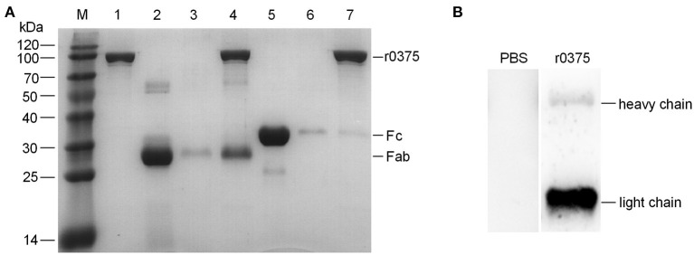
The binding region of IgG and r0375. (A) SDS-PAGE identifies that r0375 binds to the Fab fragment of human IgG. M, Molecular mass marker; Lane 1, Nickel agarose conjugated r0375; Lane 2, Fab fragment; Lane 3, Nickel agarose incubated with the Fab fragment; Lane 4, Nickel agarose conjugated r0375 incubated with the Fab fragment; Lane 5, Fc fragment; Lane 6, Nickel agarose incubated with the Fc fragment; Lane 7, Nickel agarose conjugated r0375 incubated with the Fc fragment. (B) Western blotting identifies that r0375 can bind to the light chain of IgG. Light chains and heavy chains of bovine IgG were separated by SDS-PAGE, transferred to PVDF and then incubated with PBS or r0375. The binding position was detected with mouse anti-His tag mAb and HRP-conjugated goat anti-mouse IgG.

