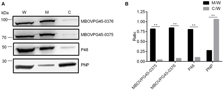
Figure 6.
Subcellular localization of MBOVPG45-0375 and MBOVPG45-0376 in M. bovis. (A) Western blotting analyzes the subcellular localization of MBOVPG45-0375 and MBOVPG45-0376. W, whole proteins of M. bovis strain PG45; M, membrane proteins; C, cytoplasmic proteins. (B) Graph showing the ratio of the protein amount in the membrane or cytoplasmic fractions to the total proteins in whole-cell lysate. The asterisk above the charts stands for statistically significant differences. **P ≤ 0.01.

