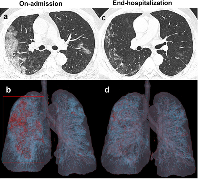
Figure 1.
Unenhanced chest CT images of a 41 years old patient infected with COVID-19 from the NF group. (a,b) Axial images obtained on-admission show extensive ground-glass opacities (GGO) in the subpleural regions of both lungs. Three-dimensional volume-rendered reconstruction (3D-VR) image mainly presents as a red lesion in the right lung (red box). (c,d) Follow-up CT images obtained at end- hospitalization show significant absorption of GGO with irregular linear opacities.

