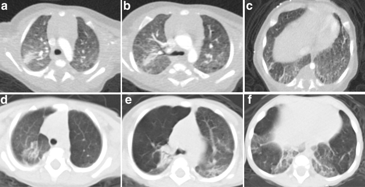
FIG. 1.
Patient 7: Axial chest CT images at 4 months of age in the upper (a), middle (b), and lower (c) lung zones demonstrate right upper lobe atelectasis, mild diffuse ground glass opacities, and basilar predominant septal thickening. Axial chest CT images at 9 months of age in the upper (d), middle (e), and lower (f) lung zones demonstrate progressive hyperinflation of right upper, left upper, and left lower lobes with persistent atelectasis and basilar septal thickening. CT, computed tomography.

