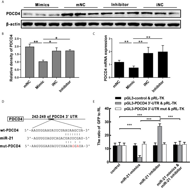
Figure 5.
Effect of miR-21 on the expression of PDCD4 in vitro. (A) HNEpC was transfected with miR-21 mimics (with mNC as control) and inhibitor (with iNC as control) for 24h. PDCD4 protein expression was determined by WB, normalized to β-actin. (B) Relative PDCD4 protein expression was quantified by densitometry based on immunoblot images. (C) PDCD4 mRNA levels were measured by qPCR. (D) The predicted miR-21 binding sites within the 3′UTR of PDCD4 mRNA. (E) Double luciferase activity assay of HEK 293 cells. After being co-transfected with the analogue of miR-21: control/miR-21 mimic/miR-21 inhibitor, and the following plasmids: pGL3-3′-UTR of control/WT/mutated PDCD4 vector and the pRL-TK vector, the ratio of GFP to RL was determined. Data were obtained in three independent experiments. One-way ANOVA was used to analyze the difference between multiple groups. The asterisk indicates statistical significance, *P < 0.05; **P < 0.01; ***P < 0.001.

