Figure 1.
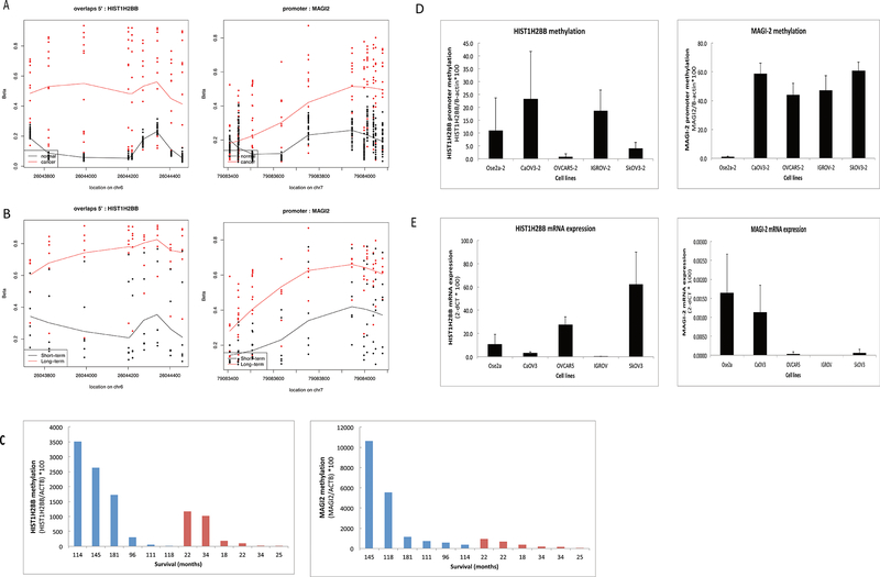
HIST1H2BB and MAGI-2 are differentially methylated in HGSC as compared to normal fallopian tube epithelium; and in HGSCs of patients with long-term survival as compared to short term survival. DNA was extracted, bisulfite-converted and analyzed by 450 K Methylation arrays and qMSP. A) Dot plots of methylation levels (Beta-values) on promoter CpG sites of HIST1H2BB and MAGI-2 in 30 normal fallopian tube samples (red) as compared to 12 HGSC samples (black). B) Dot plots of methylation levels (Beta-values) on promoter CpG sites of HIST1H2BB and MAGI-2 in 6 HGSC samples with long-term survival (red) as compared to 6 HGSC samples with short-term survival (black). C) qMSP analysis of DNA from 6 HGSC tissue samples of long-term survivors (blue) as compared to 6 HGSC tissue samples of short-term survivors (red), using primers to promoter CpG sites of HIST1H2BB or MAGI-2. Overall survival time in months is shown. D) qMSP analysis of DNA from ovarian cancer cells and a normal ovarian cell line (Ose2a), using primers to promoter CpG sites of HIST1H2BB or MAGI-2. E) RT-PCR analysis in ovarian cancer cells and a normal ovarian cell line, using primers to measure mRNA expression of HIST1H2BB or MAGI-2.

