. 2020 May 27;2(3):e180063. doi: 10.1148/ryai.2020180063

Figure 5b:

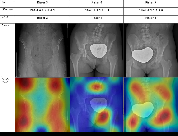
(a) Sample radiographic images graded by the automatic grading method (AGM). First row: Correctly classified. Second row: Misclassified by one grade. Third row: Misclassified by two grades. (b) Sample radiographic images. First row: ground truth (GT). Second row: Risser stage assigned by each observer. Third row: Risser stage assigned by AGM. Fourth row: Original image. Fifth row: Gradient-weighted class activation mapping (Grad-CAM) highlighting the AGM’s most important regions of images. The color map scales from red (most discriminant) to blue (least discriminant).

(a) Sample radiographic images graded by the automatic grading method (AGM). First row: Correctly classified. Second row: Misclassified by one grade. Third row: Misclassified by two grades. (b) Sample radiographic images. First row: ground truth (GT). Second row: Risser stage assigned by each observer. Third row: Risser stage assigned by AGM. Fourth row: Original image. Fifth row: Gradient-weighted class activation mapping (Grad-CAM) highlighting the AGM’s most important regions of images. The color map scales from red (most discriminant) to blue (least discriminant).