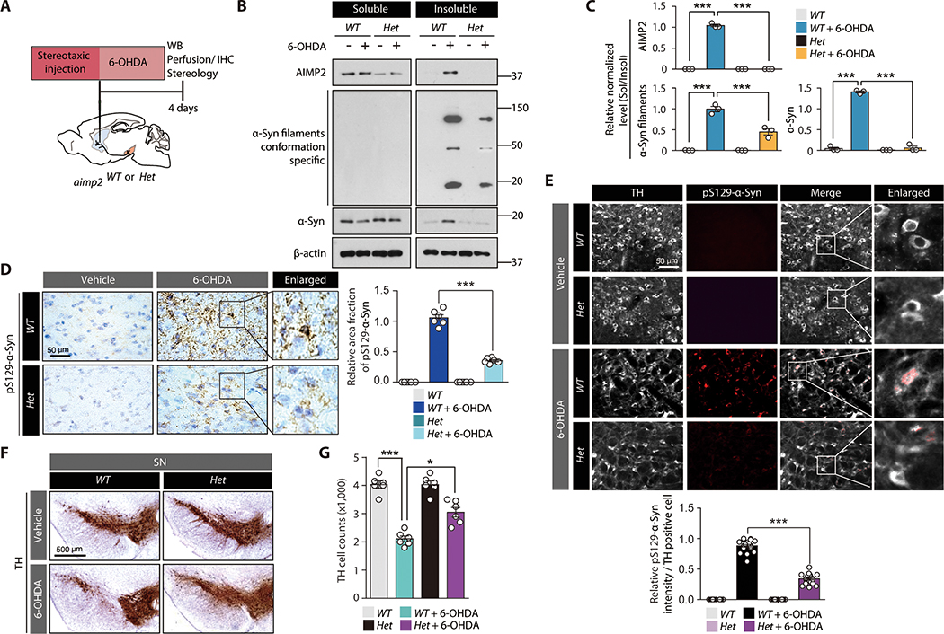
Figure 6. AIMP2 is required for 6-OHDA-induced α-synuclein aggregation and toxicity in vivo.
(A) Schematics of in vivo 6-OHDA striatal injection to 2 month old heterozygous Aimp2 KO mice (Aimp2 Het) or wild type littermate control (WT). Experimental schedules for western blot (WB), IHC, and TH stereology are indicated.
(B) Western blots of AIMP2, α-synuclein, and filament conformation α-synuclein expression in Triton X-100 soluble and insoluble fractions of the VM from the indicated experimental mouse groups.
(C) Quantification of relative amounts of AIMP2, α-synuclein, and filament conformation α-synuclein expression in insoluble fractions normalized to those in soluble fractions from sample groups in panel B (n = 3 mice per group).
(D) Neuropathological assessment of phosphorylated α-synuclein-positive inclusions in mouse brains of each experimental group monitored by immunohistochemistry using pS129 α-synuclein antibody and Nissl counter staining. Quantification of pS129-α-Syn immunohistochemistry (n = 6 slides from 3 mice per group, right panel).
(E) Representative immunofluorescence images showing pS129-α-synuclein and TH (pseudo-colored for original green fluorescence) expression in the SN of each experimental mouse group. Quantification of pS129-α-Syn immunofluorescence in TH-positive cells (n = 20 cells from 3 mice per group, bottom panel).
(F) TH immunohistochemistry images of SN sections from the indicated experimental mouse groups.
(G) Stereological counting of dopamine neurons in the SN pars compacta of each experimental mouse group (n = 5~6 mice per group).
Quantified data are expressed as mean ± SEM. Statistical significance was determined by ANOVA test with Tukey post-hoc analysis, *p < 0.05, and ***p < 0.001.

