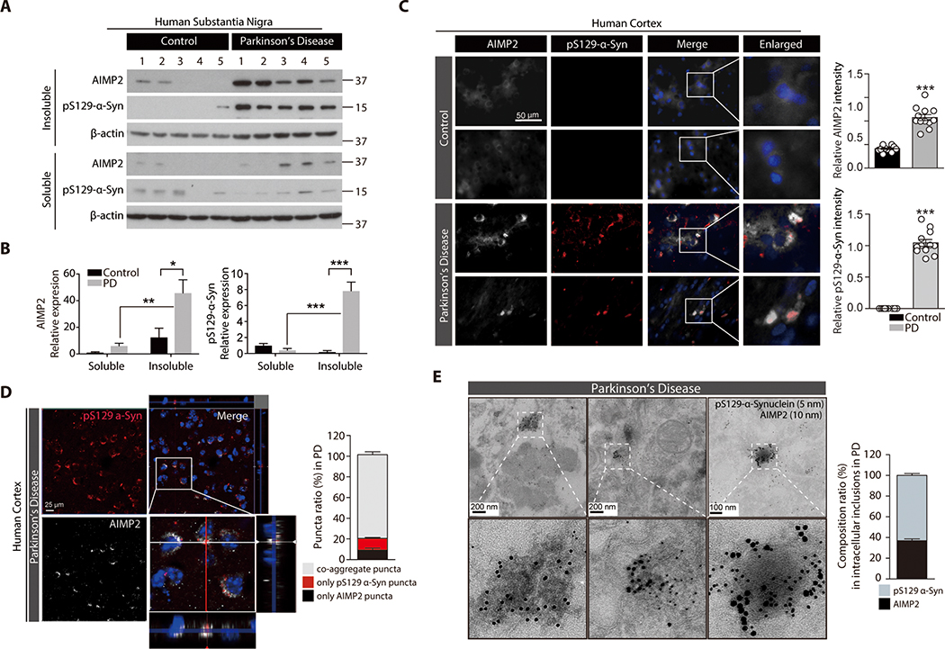
Figure 7. Insoluble co-aggregation of α-synuclein with AIMP2 in human PD brains.
(A) Distribution of AIMP2 and phosphorylated α-synuclein (pS129-α-syn) in NP-40 soluble and insoluble fractions prepared from postmortem human brains of patients with PD and age matched controls based on Western blot analysis.
(B) Relative distribution of AIMP2 and phosphorylated α-synuclein (pS129-α-syn) into each fractions normalized to β-actin (n = 5 per group).
(C) Co-aggregation of AIMP2 (pseudo-colored for original green fluorescence) into LB inclusions in temporal lobe from postmortem PD brains monitored by immunofluorescence. Quantification of AIMP2 and pS129-α-Syn immunofluorescence (n = 12 slides from 4 Con and 12 slides from 4 PD, right panel).
(D) Confocal microscopic and Z-stack images showing colocalization of AIMP2 (pseudo-colored for original green fluorescence) in the pS129 α-synuclein-positive LB in PD human brain samples. % puncta ratio composed of AIMP2 or/and phosphorylated α-synuclein (n = 21 cells from 4 PD, right panel)
(E) Ultrastructure of inclusion and colabeling of AIMP2 and pS129-α-synuclein by nanogold particles for postmortem human PD brain tissues determined by an immunoGold EM. 10 nm and 5 nm nanogold particles were used to detect anti-AIMP2 and anti-pS129-α-synuclein antibodies, respectively. Composition ratio of AIMP2 and pS129-α-Syn in inclusion structures (n = 26 inclusions from 4 PD, right panel).
Quantified data are expressed as mean ± SEM. Statistical significance was determined by ANOVA test with Tukey post-hoc analysis or unpaired two-tailed Student’s t-test, *p < 0.05, **p < 0.01, and ***p < 0.001.

