Abstract
Ipsilateral avid axillary lymph node uptake at FDG PET/CT persists in 29% (49 of 169) of patients between 7 to 10 weeks after the second dose of the mRNA-based BNT162b2 COVID-19 vaccination.
SUMMARY STATEMENT
Ipsilateral avid axillary lymph node uptake at FDG PET/CT persists in 29% (49 of 169) of patients between 7 to 10 weeks after the second dose of the mRNA-based BNT162b2 COVID-19 vaccination.
Introduction
As the novel coronavirus disease 2019 (COVID-19) pandemic crosses the one-year threshold, a growing number of countries are engaged in extended large-scale COVID-19 vaccination programs.
Recently, there have been reports of unilateral axillary lymphadenopathy seen on various imaging modalities, in association with recent ipsilateral vaccination [1,2]. A recent study estimated the prevalence of FDG PET/CT axillary lymph node uptake to be 54% up to three weeks after a second mRNA-based vaccination dose [3].
Recommendations for post vaccine lymphadenopathy seen on imaging were published, advising scheduling routine imaging, such as screening, before, or at least 6 weeks after the final vaccination dose, to eliminate false positive results [4, 5]. However, information is still lacking regarding duration and prevalence of post-vaccination imaging findings. We aimed to assess the prevalence of FDG PET/CT avid axillary lymph nodes beyond 6 weeks after the second mRNA-based BNT162b2 COVID-19 vaccine dose.
Materials and Methods
We conducted a retrospective analysis of prospectively collected data in a single tertiary medical center. The study has been approved by the institutional ethics committee, and the need for patient informed consent was waived.
All consecutive adult patients over 18 years old referred for FDG PET/CT at our institution, who performed the study at least 42 days after the second Pfizer-BioNTech (BNT162b2) COVID-19 vaccine dose, were evaluated for the presence of FDG-avid axillary lymphadenopathy ipsilateral to the injection site. Patients with underlying disease that was likely to involve the axilla (such as ipsilateral locally advanced breast cancer or lymphoma previously involving the axilla) and patients who were vaccinated in both arms, were excluded.
Bilateral axillary lymph node uptake was measured by three board certified radiologists (YE, ME, NC) and a nuclear medicine resident (YA). To overcome physiologic lymph node uptake, positive unilateral uptake was determined as SUVmax ratio between the ipsilateral and contralateral axillary lymph nodes above 1.5, as previously used [6]. Largest ipsilateral lymph node size was measured in short axis.
Results
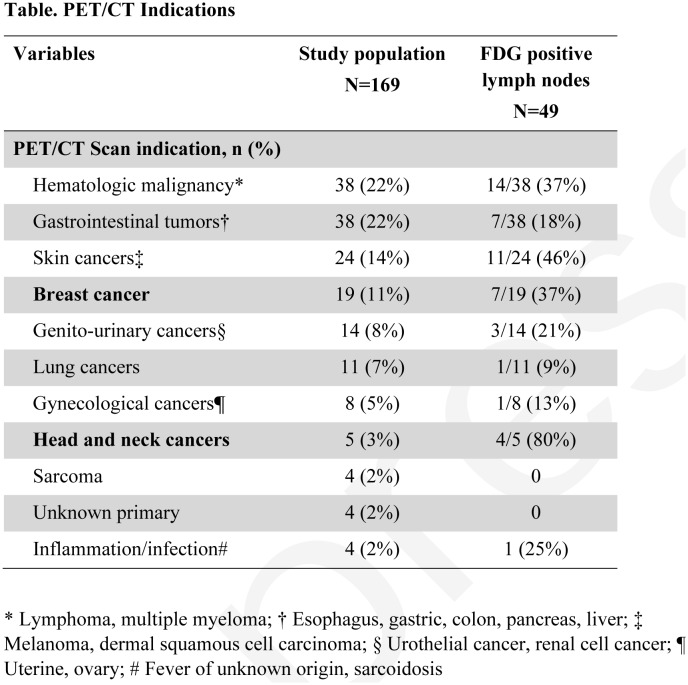
Of 205 consecutive adult subjects who had an FDG PET/CT 42 to 71 days (7–10 weeks) after a second dose vaccination, 37 were excluded: 21 had high probability of ipsilateral axillary lymph node involvement, one patient had only partial medical history information and 15 patients had bilateral injections. Our final cohort comprised a total of 169 patients (median age, 65±14 years; 49% female), scanned a median of 52±7 days after the second vaccine dose (IQR 47–57). The Table details PET/CT indications.
Table.
PET/CT Indications
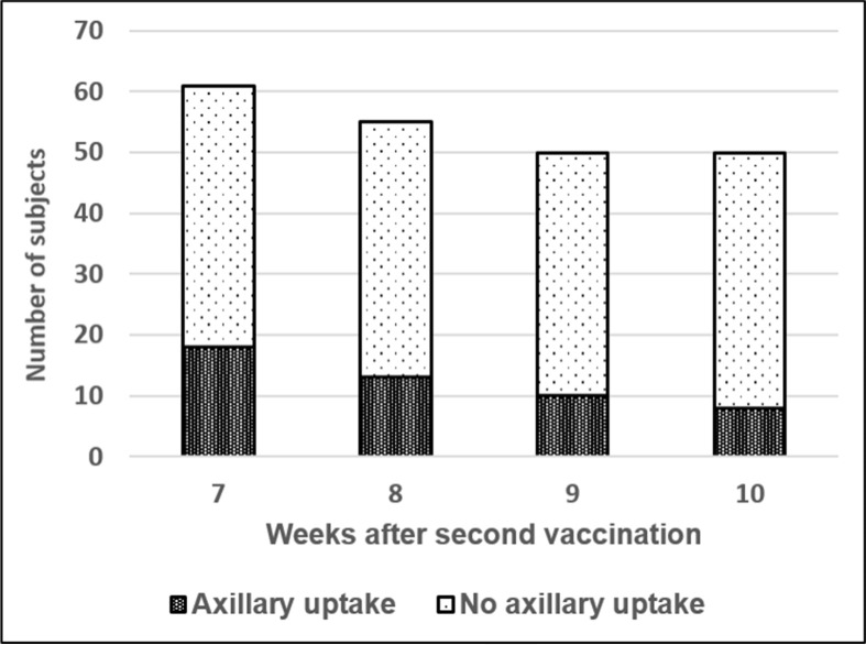
Overall, 29% (49/169) had positive axillary uptake 7–10 weeks after second vaccination (median SUV max, 2.9±1.3), divided to 42%, 31%, 25% and 19% on 7th, 8th, 9th and 10th weeks respectively. The distribution number of patients with positive uptake on each week after vaccination can be seen in Figure 1. Immunotherapy did not contribute to persistent immune reactions, as 4/14 (28%) patients receiving immunotherapy had positive lymph node uptake, compared with 13/70 (19%) patients receiving other treatment, and 32/85 (38%) patients not receiving oncologic treatment.
Figure 1:
Axillary lymph node uptake prevalence by week after second vaccination dose. Darker area corresponds to positive axillary uptake, while brighter area to negative uptake.
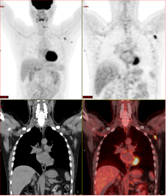
Most FDG avid lymph nodes were of normal size (mean 0.5cm, range 0.1–1.6 cm). Figure 2 depicts an example of avid axillary FDG uptake in a patient 62 days after vaccination.
Figure 2:
A 63-year-old multiple myeloma patient, with skeletal pain. New FDG avid axillary lymphadenopathy 62 days (9 weeks) after second mRNA vaccination dose.
Discussion
This study shows that avid axillary lymph node uptake on FDG PET/CT can be detected in more than a quarter of our patient population even beyond 6 weeks after the second dose of the mRNA-based COVID-19 vaccination. Compared to a previous study showing normalization of FDG uptake within 40 days of receiving an inactivated H1N1 influenza vaccine [6], we found uptake persistence even at 70 days. Physicians should be aware of this potential pitfall.
Footnotes
The authors declare no conflict of interest related to this study.
Contributor Information
Yael Eshet, Email: yael.eshet@gmail.com.
Noam Tau, Email: taunoam@gmail.com.
Yousef Alhoubani, Email: Yousef.Alhoubani@sheba.health.gov.il.
Nayroz Kanana, Email: Nayruz.Knaana@sheba.health.gov.il.
Liran Domachevsky, Email: Liran.Domachevsky@sheba.health.gov.il.
Michal Eifer, Email: michaleifer@gmail.com.
References
- 1.Eifer M, Eshet Y. Imaging of COVID-19 Vaccination at FDG PET/CT. Radiology. 2021 January 28:210030. [DOI] [PMC free article] [PubMed] [Google Scholar]
- 2.Özütemiz C, Krystosek LA, Church AL, et al. Lymphadenopathy in COVID-19 Vaccine Recipients: Diagnostic Dilemma in Oncology Patients. Radiology. 2021 February 24:210275. [DOI] [PMC free article] [PubMed] [Google Scholar]
- 3.Cohen D, Krauthammer SH, Wolf I, Even-Sapir E. Hypermetabolic lymphadenopathy following administration of BNT162b2 mRNA Covid-19 vaccine: incidence assessed by [18F]FDG PET-CT and relevance to study interpretation. Eur J Nucl Med Mol Imaging. 2021 March 27. [DOI] [PMC free article] [PubMed] [Google Scholar]
- 4.Becker AS, Perez-Johnston R, Chikarmane SA, Chen MM, El Homsi M, Feigin KN, Gallagher KM, Hanna EY, Hicks M, Ilica AT, Mayer EL, Shinagare AB, Yeh R, Mayerhoefer ME, Hricak H, Vargas HA. Multidisciplinary Recommendations Regarding Post-Vaccine Adenopathy and Radiologic Imaging: Radiology Scientific Expert Panel. Radiology. 2021 February 24:210436. [DOI] [PMC free article] [PubMed] [Google Scholar]
- 5.Tu W, Gierada DS, Joe BN. COVID-19 Vaccination-Related Lymphadenopathy: What To Be Aware Of. Radiology, Imaging Cancer Published Online:Apr 9 2021 https://doi.org/10.1148/rycan.2021210038 [DOI] [PMC free article] [PubMed] [Google Scholar]
- 6.Thomassen A, Lerberg Nielsen A, Gerke O, Johansen A, Petersen H. Duration of 18F-FDG avidity in lymph nodes after pandemic H1N1v and seasonal influenza vaccination. Eur J Nucl Med Mol Imaging. 2011 May;38(5):894–8. [DOI] [PubMed] [Google Scholar]


