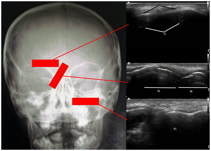
Figure 1.
Sinus ultrasound protocol: Left image the transducer positioning, upper right image at the level of the frontal sinus; middle right image at the level of the lateral aspect of the nasal pyramid; lower right image at the level of the maxillary sinus. Therefore, one can visualize the anterior wall of the sinus 1) and the posterior echo generated through the encounter of ultrasound rays with the posterior sinus wall 2). The structures visualized are nasal bones 3) with posterior shadow effect and alar cartilages 4). In addition, the integrity of the anterior sinus wall 5) and the posterior back echo generated at the level of the posterior wall of the sinus 6) are visualized.

