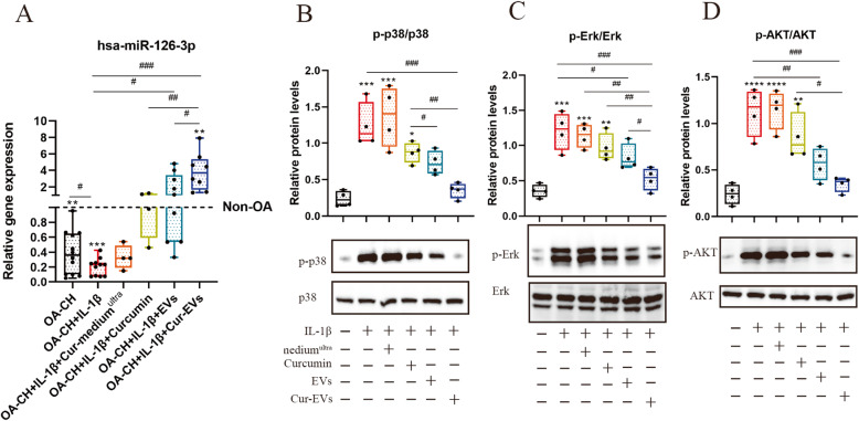
Fig. 8.
Effect of Cur-EVs on phosphorylation of Erk1/2, PI3K/Akt, and p38 MAPK in IL-1β-treated OA-CH and on gene expression of hsa-miR-126-3p. Phosphorylation levels of Erk1/2, PI3K/Akt, and p38 MAPK in OA-CH were detected by western blotting. a Gene expression of hsa-miR-126-3p in OA-CH, OA-CH, IL-1β-induced OA-CH in different treatment groups (Cur-mediumultra, free curcumin (10 μM), EVs and Cur-EVs). Difference to non-OA-CH (dotted line); **p < 0.01; ***p < 0.001, #difference between groups: #p < 0,05; ##p < 0,01; ###p < 0,001; 1-way ANOVA with Newman-Keuls multiple comparison test; n = 4–15. b–d Representative western blot images and quantification of phosphorylation level of Erk1/2, P13K/Akt, and p38 MAPK after densitometric analysis. All values represent mean ± standard deviation. *Compared with no treatment control (OA-CH), *p < 0.05; **p < 0.01; ***p < 0.001; ****p < 0.0001; #difference between groups, #p < 0,05; ##p < 0,01; ###p < 0,001; one-way ANOVA with Newman-Keuls multiple comparison test; n = 4

