
Figure 4: Loss of the clock gene Bmal1 in Nav1.8+ sensory neurons leads to reduced OA (osteoarthritis) pain.
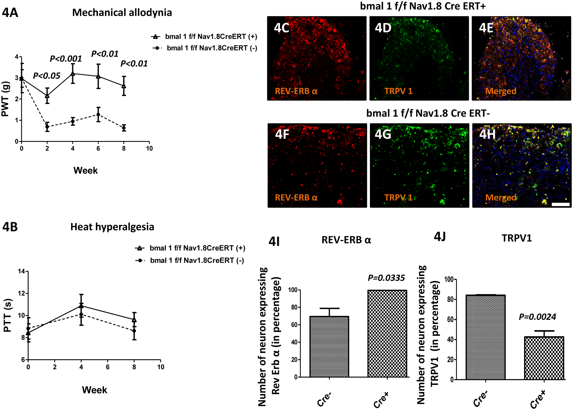
(Fig. 4A) Mechanical hyperalgesia was measured using von Frey filament test. The paw withdrawal threshold (PWT) of the bmal1f/f Nav1.8CreERT(−) group (n = 5) were low starting at 2 weeks after OA induction, and were lower than bmal1f/f Nav1.8CreERT(+) group (n = 5) at all time points. (Fig. 4B) Heat hyperalgesia was measured using hot-plate test. The paw thermal threshold (PTT) in the bmal1f/f Nav1.8CreERT(−) & bmal1f/f Nav1.8CreERT(+) group was not significantly changed. However, expression of Rev-Erb-α in the dorsal root ganglia (DRG) following OA surgery was higher and TRPV1 was lower in in bmal1f/f Nav1.8CreERT(+) vs. bmal1f/f Nav1.8CreERT(−) mice. Double immunofluorescence staining of Rev-Erb-α (red; Fig. 4C and Fig. 4F) and TRPV1 (green; Fig. 4D & Fig. 4G) in DRGs of bmal1f/f Nav1.8CreERT(−) & bmal1f/f Nav1.8CreERT(+) groups mice. Co-localization of the two stains appears yellow (Fig. Fig. 4E & Fig. 4H). A scale bar was 20μm and is shown in white in color for all images. Quantitative analyses of Rev-Erb-α and TRPV1 expression in DRG (Fig. 4I-4J). Values are mean±SEM.