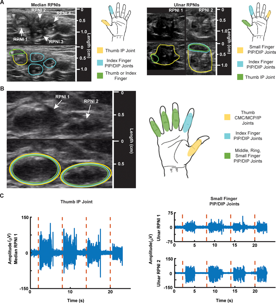
Fig. 2. RPNI sonograms, motor map, and electrophysiology.
(A) P1’s median and ulnar RPNI sonograms captured 19 months after RPNI surgery. Encircled areas on the sonogram show which region of the median or ulnar RPNIs contracted during cued finger movements. (B) P2’s sonogram of two RPNIs captured 8 months after RPNI surgery and motor map of active areas. (C) P1’s EMG signals (blue) recorded from median RPNI 1 after cued thumb IP joint movement (red dashed line), and EMG signals (blue) recorded from ulnar RPNI 1 and RPNI 2 after cued small finger PIP/DIP movement (red dashed line).

