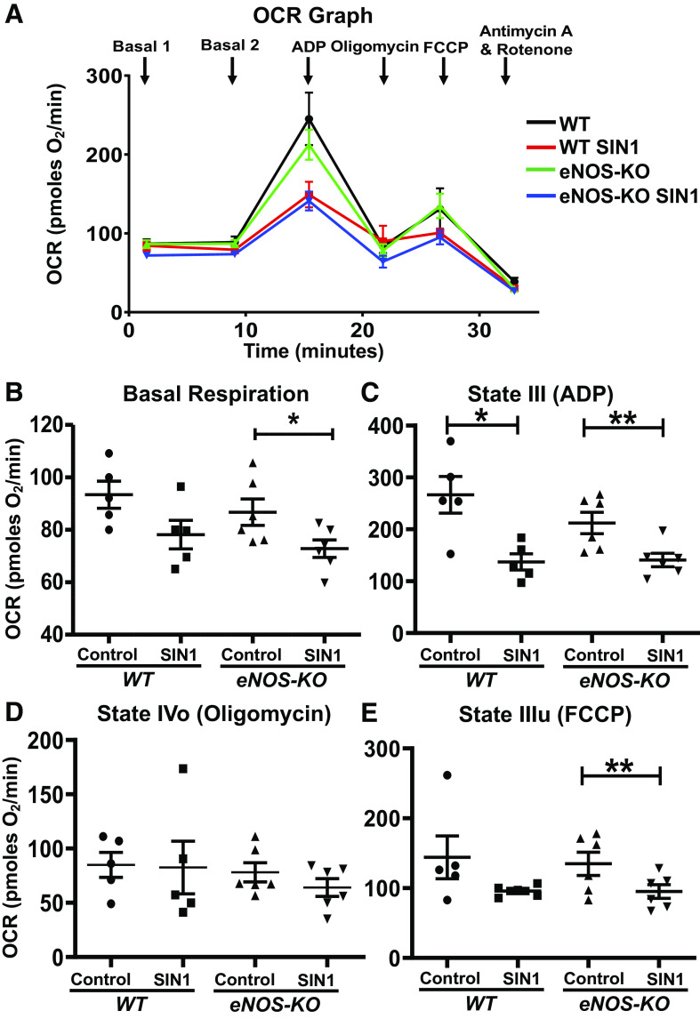
Figure 2.
SIN-1-1 (50 µmol/L) decreases mitochondrial respiration in isolated brain mitochondria. A: oxygen consumption rate (OCR) versus time graph. B basal or state II respiration. C: state III respiration (ADP-induced). D: state IVo respiration (oligomycin-inhibited). E: state IIIu [carbonyl cyanide-4-(trifluoromethoxy)phenylhydrazone (FCCP)-induced]. Mitochondria were isolated from wild-type (WT; n = 5 mice) and endothelial NO synthase knockout (eNOS-KO) mice (n = 6 mice) brains, treated with SIN-1 (50 µmol/L), and respiratory parameters were measured using Agilent Seahorse XFe24 analyzer. Data are presented as means ± SE and analyzed by repeated-measures two-way ANOVA (data were log-transformed for some parameters). *P < 0.05 and **P < 0.01, statistically significant.

