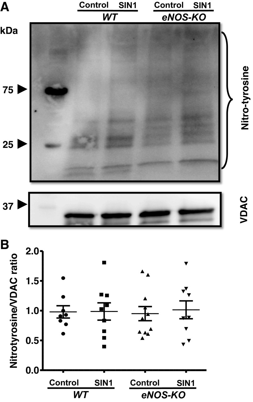
Figure 5.
SIN-1 (50 µmol/L) has no significant effect on nitrotyrosination of mitochondrial proteins. A: representative Western blot for nitrotyrosine content and VDAC (loading control). B: representative bar diagram. Mitochondria were isolated from wild-type (WT) and endothelial NO synthase knockout (eNOS-KO) mice (n = 8–11 mice) brains, treated with SIN-1 (50 µmol/L). Mitochondrial pellets were solubilized with NP 40 buffer and subjected to SDS-PAGE (4%–20% gradient). Nitrotyrosine content in the proteins was detected on Immun-Blot using enhanced chemiluminescence. Total band intensity was measured using ImageJ. Data are presented as means ± SE and analyzed by repeated-measures two-way ANOVA.

